The sample pattern will depend on what type of biopsy gun is used. For a standard spring-loaded gun, five samples should be obtained in the 12, 3, 6, and 9 o’clock positions for masses. For calcifications, Langer advised removing five samples in the same formation and then doing a specimen radiograph. If the calcifications do not appear in the specimen, repeat the process until a maximum of 12 to 15 samples are obtained.
For vacuum-assisted biopsy, Langer said he removes about eight tissue samples from the mass or calcifications during one rotation of the device. He collects between 16 and 24 core samples after one to two complete revolutions. The benefit of using the vacuum-assisted technique is that it improves histological diagnosis because of the higher number of available samples.
Imaging
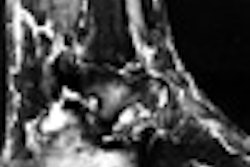
Three sets of images must be obtained for an accurate biopsy: Stereotactic images for targeting, pre-fire stereo images, and post-fire images.
"First, we’re going to start with the stereotactic image that we’re going to use for targeting," Langer said. "On these, we’d like to see the lesion with certainty. We’ll then calculate the position in three-dimensional space by targeting the center of the mass. We’d like to see the needle pointed at the lesion."
The pre-fires images are taken once the needle has been advanced. Langer called these the most important images because they should show the needle in place and aimed at the lesion.
"If these two images are positioned properly relative to the lesion, you almost can’t miss. I suppose the patient could suddenly move, but otherwise, you are going to have an accurate biopsy. Any targeting corrections that you need to make should be made on the basis of these images," he said.
Finally, the post-fire images will verify that the needle has gone through the lesion.
Targeting correction
If pre-fire images show that the needle is not aligned with the lesion, repositioning will be in order, which requires thinking back to "terrible stuff from 11th grade geometry," Langer said. That’s because the two main biopsy devices use two different calculation systems.
The Lorad machine (Trex Medical of Danbury, CT) uses Cartesian coordinates, which are more intuitive, Langer said. "The x-axis is always parallel to the floor. The y-axis is always perpendicular to the x and the z-axis is always into the patient," he explained. "The location of any lesion in the breast, at any point in space, can be defined by its location in these three planes."
The Fischer Imaging system (Denver) works with polar coordinates. In this system, the position of the lesions depends on the radius vector or the length of the needle. Then two angles help determine the exact position: The vertical angle ("Is the lesion above or below the radius vector?" and the horizontal angle ("Is it to the left or the right of the radius vector?").
"You have to move the needle in terms of the angles, and this is a little bit more complicated," Langer said. "We are dealing with the two angles. The distance that something moves in the breast is related to these angles, but it’s also related to the length of the needle."
While there is no universal formula for figuring out the vector, Langer did offer one shortcut: For the 11-gauge vacuum-assisted Mammotome needle, a change in angle of 0.19° equals an axis change of 1 mm.
"You can ignore which technique they use if the needle winds up right on target," he said. "But if you have to make a position correction, you have to understand about the two different techniques."
By Shalmali Pal
AuntMinnie.com staff writer
October 10, 2000
1 2
Let AuntMinnie.com know what you think about this story.
Copyright © 2000 AuntMinnie.com