

AuntMinnie
  • Europe
  • Clinical News
  • Informatics
  • Industry News
  • Practice Management
  • Education
  • Subspecialties
  • More
  • Sign In
  • Board Review
  • CME
  • Careers
  • Cases
  • Conferences
  • Videos
  • Webinars
  • Podcast Network
  • Advertising
  • Buyer's Guide
  • Vendors
  • Minnies
  • RPT Tracker
Clinical News
  • CT
  • Digital X-Ray
  • Interventional
  • Molecular Imaging
  • MRI
  • Radiation Oncology/Therapy
  • Ultrasound
  • Womens Imaging
Imaging Informatics
  • Advanced Visualization
  • Enterprise Imaging
  • AI
  • Cybersecurity
Industry News
  • Installations
  • Product News
  • Regulatory
  • M&A
  • Market Analysis
Practice Management
  • Administration
  • Associations
  • Careers
  • Equity & Inclusion
  • Legislation
  • Medicolegal
  • Patient Safety
  • Radiologic Technologist
  • Service
  • Salary Scan
Resources
  • Board Review
  • CME
  • Careers
  • Cases
  • Conferences
  • Videos
  • Webinars
  • Podcast Network
  • Advertising
  • Buyer's Guide
  • Vendors
  • Minnies
  • RPT Tracker
User Tools
  • Sign In
More
  • About Us
  • Advertising
  • Help
Follow AuntMinnie
Facebook iconTwitter X icon LinkedIn iconInstagram icon
  • Europe
  • Clinical News
    • CT
    • Digital X-Ray
    • Interventional
    • Molecular Imaging
    • MRI
    • Radiation Oncology/Therapy
    • Ultrasound
    • Womens Imaging
  • Informatics
    • Advanced Visualization
    • Enterprise Imaging
    • AI
    • Cybersecurity
  • Industry News
    • Installations
    • Product News
    • Regulatory
    • M&A
    • Market Analysis
  • Practice Management
    • Administration
    • Associations
    • Careers
    • Equity & Inclusion
    • Legislation
    • Medicolegal
    • Patient Safety
    • Radiologic Technologist
    • Service
    • Salary Scan
  • Education
    • Medical Students
    • Residents/Fellows
  • Subspecialties
    • Breast Imaging
    • CV
    • Chest
    • Emergency
    • GI
    • GU
    • Head & Neck
    • Interventional
    • Physics
    • MSK
    • Neuro
    • Nuclear
    • Pediatric
    • Radiation Oncology
  • More
  • Board Review
  • CME
  • Careers
  • Cases
  • Conferences
  • Videos
  • Webinars
  • Podcast Network
  • Advertising
  • Buyer's Guide
  • Vendors
  • Minnies
  • RPT Tracker
  • Sign In
Follow AuntMinnie
Facebook iconTwitter X icon LinkedIn iconInstagram icon

Will Morton

[email protected]
CT
Stepped reminders boost CT lung cancer screening
Trial findings directly address the current lack of effective lung cancer screening interventions.
August 11, 2026
Lung Cancer
Practice Management
Patient-friendly radiology reports improve family comprehension
Terminology in radiology reports can be difficult for patients and families to comprehend.
August 5, 2026
Pediatric Imaging
Interventional
CT-selected thrombectomy shows 1-year benefit in large-core stroke
Notably, the TESLA trial used a noncontrast CT (NCCT)-alone patient selection protocol.
August 3, 2026
Aneurysm
Molecular Imaging
PET/MRI detects arterial inflammation in prostate cancer patients
Androgen deprivation therapy (ADT) is widely used in advanced prostate cancer, yet is associated with increased cardiovascular events such as stroke and heart attacks.
July 31, 2026
Representative axial fused F-18 FDG-PET/MRI images and corresponding anatomic MRI images from a patient imaged at baseline and after initiation of androgen deprivation therapy are shown. Each row displays a distinct vascular territory. White arrows indicate focal areas of increased tracer uptake within the arterial wall, where spherical volumes of interest (VOIs) were placed for maximum standardized uptake value (SUVmax) measurement. Additional VOIs were placed in the superior vena cava and inferior vena cava for blood-pool activity sampling. SUVmax values from the arterial VOIs were extracted on the corresponding PET images and normalized to the blood-pool mean standardized uptake value to derive the maximum target-to-background ratio.
CT
Nongated CT scans can predict heart disease risk
Subjects with moderate and severe CAC scores on nongated CT had significantly higher risk of coronary heart disease risk.
July 30, 2026
Heart 1
CT
CT radiation doses have fallen sharply over past decade
The largest reductions appeared in chest examinations with contrast material, where diagnostic reference levels fell 31%.
July 28, 2026
Bar graphs show comparison of volume CT dose index (CTDIvol) (A) achievable doses (50th percentile) and (B) diagnostic reference levels (75th percentile) between 2014 (see reference 4 by Kanal et al) and 2025 (present study) for 10 common CT examination types performed without contrast (w/o) and with contrast (w/). Across all 10 adult CT examination categories, CTDIvol achievable doses and diagnostic reference levels generally decreased from 2014 to 2025, with the largest reductions for torso examinations (chest; abdomen-pelvis; and combined chest, abdomen, and pelvis), intermediate reductions for neck and cervical spine, and minimal change for head examinations.
Molecular Imaging
SPECT/CT viable over PET/CT for high-risk prostate cancer staging
Considering its broader accessibility, Tc-99m MIP-1404 CZT SPECT/CT may represent a practical alternative for primary staging.
July 24, 2026
Maximum intensity projection of Ga-68 PSMA-11 PET/CE-CT (A), Tc-99m MIP-1404 CZT SPECT/CT (B) and Tc-99m HDP CZT SPECT/CT (C) imaging. Ga-68 PSMA-11 PET/CE-CT and Tc-99m MIP-1404 CZT SPECT/CT showing tracer uptake in lymph nodes below the iliac bifurcation and above, paraaortic and left supraclavicular, bone uptake in the right proximal femur and right scapula (arrows). Tc-99m HDP CZT SPECT/CT did not show any uptake suspicious for metastatic disease. No sclerotic lesions suspicious of metastases were detected by the CT component in any of the studies. Fusion axial imaging of Ga-68 PSMA-11 PET/CE-CT (D) and Tc-99m MIP-1404 CZT SPECT/CT (E) showing uptake in lymph nodes in the left pelvic area below the iliac bifurcation (arrows). Fusion axial imaging of Ga-68 PSMA-11 PET/CE-CT (F) and Tc-99m MIP-1404 CZT SPECT/CT (G) showing uptake in the right proximal femur, not detected by Tc-99m HDP CZT SPECT/CT (H), (arrows).
Editor's Note
AuntMinnie.com Molecular Imaging Insider
In this edition of the Molecular Imaging Insider, we’ve highlighted a study showing that Lutetium-177 PSMA-617 spots early signs of benefit in men with metastatic hormone-sensitive prostate cancer.
July 21, 2026
Nuclear Medicine
Lutetium-177 PSMA-617 shows promise in earlier-stage prostate cancer
Across 14 studies, PSA50 response rates (the number of patients with at least a 50% drop in PSA) ranged from 44% to 100%.
July 21, 2026
Prostate Imaging
CT
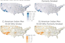
LDCT screening criteria miss majority of Korean lung cancer patients
Just 35.4% of patients diagnosed with lung cancer met the 2023 American Cancer Society (ACS) screening criteria.
July 21, 2026
Lung Cancer
Previous Page
Page 2 of 57
Next Page
Top Stories
Examples of ultrasound findings and techniques. (A) Images in a 39-year-old male patient with a mass in the left thigh. The mass is heterogeneous on the B-mode US image (compared with the patient in D) and showed increased microvascularity (superb microvascular imaging [SMI]) and shear-wave elastography (SWE) values. Undifferentiated pleomorphic sarcoma was diagnosed at biopsy (with pleomorphic rhabdomyosarcoma in surgical specimen). (B) Images in an 18-year-old male patient with a mass in the left leg. The mass is hypoechoic on the B-mode image, with no other findings suggestive of malignancy. The lesion is in contact with the cortex of the tibia, which is slightly irregular. CT revealed a doubtful anteromedial tibial erosion. The microvascular study demonstrated high vascularization, suggestive of malignancy. Periosteal Ewing sarcoma was diagnosed with both histologic and immunohistochemical confirmation. (C) Images in a 69-year-old female patient with a lump growing on the outside of the left leg. Multiple SWE examinations were performed (please note the high values obtained in the measurements, whereas the color map highlights the stiffness relative to adjacent tissues). SMI showed areas of increased vascularization to target for sampling. Undifferentiated spindle cell sarcoma was diagnosed at biopsy, with residual leiomyosarcoma in the surgical specimen after neoadjuvant therapy. (D) Images in a 56-year-old female patient with a mass in the right thigh. The mass is heterogeneous at both B-mode ultrasound (similar to patient A) and MRI (coronal T2-weighted spectral attenuated inversion recovery [SPAIR]; T1-weighted pre-contrast and postcontrast imaging), which even shows uptake after the administration of paramagnetic contrast material, which is traditionally suggestive of malignancy. Low values at SMI and elastography are suggestive of benignity. Spindle cell lipoma was diagnosed at biopsy, with atypical spindle cell lipomatous tumor in the surgical specimen.
Ultrasound
Advanced ultrasound techniques improve soft tissue tumor evaluation
Both SWE and SMI can help distinguish benign from malignant soft tissue tumors.
Woman Patient Asian 400
Womens Imaging
Should cancer data stop lumping Asian Americans, NHPI people together?
Serial axial chest CT scans demonstrating the longitudinal evolution of small pure ground-glass nodules (pGGNs) incidentally detected during a routine wellness assessment. (A) Images in a 37-year-old woman with a family history of lung cancer show a nodule with the progressive growth pattern (box). The baseline CT scan, obtained in October 2017, demonstrates a pure ground-glass nodules in the right lower lobe. Consecutive CT scans obtained from 2017 to 2022 show the continuous enlargement of the nodule during active surveillance. The maximum diameter of the nodule increased from 5.6 mm in 2017 to 10.2 mm in 2022. (B) Images in a 45-year-old man show a nodule with the indolent growth pattern (box). The baseline CT scan, obtained in April 2018, demonstrates a pure ground-glass nodules in the left lower lobe. Consecutive CT scans obtained from 2019 to 2023 show that the nodule remained radiographically stable during CT surveillance. The maximum diameter of the nodule was 5.4 mm in 2018 and 6.1 mm at the final surveillance scan in 2023.
CT
CT reveals progressive growth pattern in incidental lung nodules
Shutterstock 1865403334
Practice Management
Radiology faculty most stressed, least satisfied
Nominations Are Open — 2026 Minnies
Sponsor Content
Nominations Are Open — 2026 Minnies
More in Home
Radiotherapy access could address gynecological cancer disparities
By Amerigo Allegretto
Inadequate radiotherapy infrastructure is one of several drivers of gynecological cancer disparities in LMICs.
August 24, 2026
Radiotherapy 400
Food insecurity linked to higher risk of death in breast cancer
By Will Morton
Food-insecure regions are where people lack consistent access to enough affordable, nutritious food.
August 21, 2026
Food Insecurity
How much surveillance imaging is needed for meningiomas?
By Amerigo Allegretto
Most surveillance MRI or CT exams did not result in intervention, and treatment rates remained stable during the study period.
August 21, 2026
Mri Slide 400
Ultrasound MinnieCast, Episode 15: Wearable ultrasound making strides
By Amerigo Allegretto
Geonho (Tom) Park, PhD, from Stanford Medicine shares insights from the work he and colleagues have done in wearable ultrasound.
August 21, 2026
Minnie Cast Thumbnail (20)
PET/CT outperforms MRI after focal therapy for prostate cancer
By Will Morton
No accurate noninvasive method has been validated to evaluate treatment response after focal therapy.
August 20, 2026
True-positive PSMA-PET with positive MRI in 74-year-old male with prostate cancer (Gleason score of 4 + 5; left anterior and posterior apex positive cores). (A) Left posterior peripheral apical to midprostate hyposignal visible on transaxial MRI. (B) No definite CT correlate evident (white arrow). (C) Focal high PSMA expression detectable in left posterior mid to base with SUVmax of 12.4 on transaxial fused delayed pelvic PET/CT (yellow arrow). (D) Focal high PSMA expression found in left midgland to base of prostate on maximum-intensity-projection image (yellow arrow).
Florida approves NBPAS as recognized board certification option
By AuntMinnie.com staff writers
NBPAS-certified physicians can now represent themselves as board certified in Florida physician advertising.
August 20, 2026
Florida Sign 400
Thermal ablation achieves long-term survival in kidney cancer
By Will Morton
The 10-year overall survival (OS) and cancer-specific survival (CSS) rates were 68.5% and 75.5%.
August 20, 2026
Kidney2
Most FDA-cleared AI medical devices not tested on patient outcomes
By Amerigo Allegretto
Of the 1,059 FDA-cleared devices used in radiology, three had a registered trial.
August 19, 2026
Artificial Intelligence Ai Global Communication Network 400
AI framework could improve MRI quality control in prostate cancer
By Amerigo Allegretto
The framework uses a DL model trained on low- and high-quality MR images and helped improve radiologist accuracy.
August 19, 2026
Low-quality axial T2-weighted MRI scans with gradient-weighted class activation mapping. The low-quality MRI scans (top row) show blurred structures and artifacts making zonal and capsule boundaries indistinguishable. On the same scans with gradient-weighted class activation map overlays (bottom row), red indicates regions that were most heavily weighted by the deep learning model toward the low-quality prediction.
New immuno-PET tracer shows promise in lung cancer
By Will Morton
Molecular imaging can provide whole-body visualization and quantitative assessment of PD-L1 expression.
August 18, 2026
(A) Ga-68 NK224 PET/CT maximum intensity projection image (left), axial PET/CT images (center), and axial CT images (right) in a 43-year-old female participant with lung adenocarcinoma show lesions in the right cerebellum (blue arrow), right frontal lobe (red arrow), and sternum (green arrow) with low, intermediate, and high tracer uptake, respectively. Photomicrographs (scale bar, 100 μm) with hematoxylin-eosin (H&E) staining (center bottom) and programmed death ligand 1 (PD-L1) immunohistochemical (IHC) staining (right bottom) show high PD-L1 expression in the sternal lesion (tumor proportion score ≥ 50%).
AI fracture detection device validated across three European hospitals
By Will Morton
No differences in AI performance were observed across centers, and AI assistance was associated with higher reader sensitivity.
August 18, 2026
Artificial intelligence (AI) assistance facilitates correct fracture identification in cases initially missed by human readers. A representative case is shown using anteroposterior radiographs in a 29-year-old male patient with wrist trauma (suspected nondisplaced transverse scaphoid fracture). The reader’s initial unaided assessment (top) resulted in a missed fracture, whereas with AI assistance (bottom), the fracture was correctly identified. The green box represents the fracture location as predicted by the AI tool, the blue box represents the reference standard annotation, and the red dots represent individual reader annotations.
Radiology Partners unit Mosaic petitions FDA on imaging AI regulation
By AuntMinnie.com staff writers
The petition seeks clarity on the regulation of AI vision language models used for diagnostic medical image analysis.
August 18, 2026
Artificial Intelligence Ai Medical Healthcare Tools 400
Previous Page
Page 2 of 246
Next Page
AuntMinnie
Topics
  • Clinical News
  • Imaging Informatics
  • Industry News
  • Practice Management
  • Radiology Education
  • Subspecialties
Resources
  • Board Review
  • CME
  • Careers
  • Cases
  • Conferences
  • Videos
  • Webinars
  • Podcast Network
  • Advertising
  • Buyer's Guide
  • Vendors
  • Minnies
  • RPT Tracker
More
  • About Us
  • Advertising
  • Help
Follow AuntMinnie
Facebook iconTwitter X icon LinkedIn iconInstagram icon
  1. Privacy Policy
  2. Privacy Settings
  3. Terms & Conditions
  4. Contact Us
  5. Site Map
  6. Home
© 2026 Science and Medicine Group. All rights reserved.