Nongated CT scans can predict heart disease risk as accurately as dedicated cardiac scans, offering a scalable screening approach that could help identify high-risk patients among the 19 million chest CT scans performed annually in the United States.
- Comparable Accuracy: Nongated CT scans predict cardiovascular events with performance comparable to gated cardiac CT scans, according to a study published in Circulation.
- Risk Stratification: Participants with moderate and severe coronary artery calcium scores had significantly higher risk of coronary heart disease (hazard ratios 2.67 and 5.22) and cardiovascular disease (hazard ratios 1.32 and 2.89).
- Scalable Approach: With 19 million nongated chest CT scans performed annually in the U.S., incorporating coronary artery calcium quantification offers a cost-effective preventive care strategy.
- AI-Ready Technology: FDA-approved AI algorithms are now available to automatically identify coronary artery calcium in routine chest CT scans.
- Clinical Implementation: The 2026 ACC/AHA dyslipidemia guidelines recommend using incidental CAC from nongated CT scans to guide lipid-lowering therapy decisions.
Coronary artery calcium scores derived from routine nongated chest CT scans predict cardiovascular events with accuracy comparable to dedicated cardiac CT scans, according to a study published July 27 in Circulation.
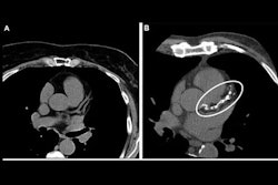
A group from Massachusetts General Hospital in Boston analyzed paired gated and nongated CT scans from a large, multiethnic U.S. cohort, with findings suggesting that nongated chest CT scans could serve to screen for cardiovascular disease in millions of patients every year.
“Given the large number of nongated scans performed annually, incorporating their quantification into clinical practice offers a scalable approach to personalized preventive care,” noted lead author Dhiran Verghese, MD, and colleagues.
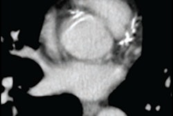
Coronary artery calcium (CAC) scoring is a well-validated predictor of atherosclerotic cardiovascular disease and is typically measured on dedicated electrocardiogram (ECG)-gated CT scans, yet CAC remains underused in clinical practice, largely because of lack of insurance coverage for ECG-gated CAC scans, the authors explained.
Conversely, CAC scores can be derived incidentally from nongated chest CT scans, of which 19 million are performed annually in the U.S. each year, often in healthy people, for example to screen for lung cancer. Nonetheless, CAC quantification is typically not undertaken on these scans due to limited data on the association of incidentally identified nongated CAC and cardiovascular outcomes, the group noted.
To bridge the gap , the researchers gathered data from 2,472 participants from a previous study who underwent same-day gated and nongated chest CT scans between April 2010 and December 2011. Scans were interpreted at a blinded core laboratory using the Agatston methodology, with CAC categorized as none (0), mild (1–99), moderate (100–299), or severe (≥300).
According to the findings, compared with participants with no CAC, those with moderate and severe CAC scores on nongated CT had significantly higher risk of coronary heart disease risk (hazard ratios [HR], 2.67 and 5.22) and cardiovascular disease risk (HR, 1.32 and 2.89). Also, nongated and gated log-standardized CAC scores were highly correlated (r = 0.961), and the area under the receiver operating characteristic curve, C-statistic, and Brier scores were statistically similar between the two scan types for all outcomes.
“Nongated CAC predicts cardiovascular events with performance comparable to gated CAC,” the researchers wrote.
The group noted that the findings are particularly relevant given the release of 2026 dyslipidemia guidelines by the American College of Cardiology/American Heart Association that recommend using incidental CAC from nongated CT scans to guide lipid-lowering therapy decisions.
In an accompanying editorial, Fatima Rodriguez, MD, and Curtis Langlotz, MD, PhD, both of Stanford University in Palo Alto, CA, noted that several AI algorithms are now U.S. Food and Drug Administration–approved and validated to complete the task.
“Systematically identifying CAC in millions of chest CTs performed each year for noncardiac purposes should be prioritized because of its potential for public health impact. This new method of CAC detection represents the promise of precision prevention at scale – leveraging existing data to identify high-risk individuals and offering tailored interventions to motivate sustained change,” they wrote.
The evidence is clear, the technology is ready, and the time for clinical implementation is now, Rodriguez and Langlotz concluded.
Read the full study here.