Dear Digital X-Ray Insider,
The scapulohumeral rhythm (SHR) -- the coordinated motion of the scapula and humerus during shoulder movement -- was first described in 1934 as a metric for shoulder function, yet there remains no current imaging method for measuring SHR that is both cost-effective and efficient for clinical use. Dynamic digital radiography (DDR) could be a game changer in this regard, according to a group at Emory University in Atlanta.
The researchers recently introduced a DDR technique that appears efficient and reproducible for measuring SHR in patients with rotator cuff tears and other shoulder injuries. You can read about the research in this edition's Insider Exclusive.
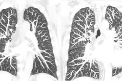
Meanwhile, other advances in digital x-ray continue apace. A group in Japan demonstrated for the first time that suppressing bone artifacts on temporal subtraction (TS) digital chest x-rays can significantly improve visual evaluation of pulmonary lesions.
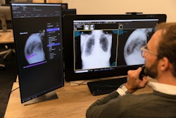
And in artificial intelligence (AI) research, several new algorithms for detecting abnormalities on chest x-rays appear promising, as illustrated in the following stories:
- A team in Korea added a bone-suppression algorithm to a deep-learning model they previously developed and found the model helped radiologists reach sensitivities of 92% for detecting lung nodules.
- Researchers in India developed an AI model for detecting active tuberculosis that could increase real-world applications of the technology in primary-care settings with limited resources.
- A team at Boston's Massachusetts General Hospital developed an AI deep-learning model that identifies patients with acute chest pain syndrome who are at risk for 30-day acute coronary syndrome, pulmonary embolism, or aortic dissection.
Also, identifying fractures using x-rays technology remains a stalwart strategy, with researchers in Hungary suggesting that bone scans may predict fracture risk in psoriatic arthritis patients and a Dutch group recommending spinal x-rays to determine the true risk of vertebral fractures in men with prostate cancer prior to androgen deprivation therapy.
In other stories we covered, fluoroscopy revealed its continued value in a case series by researchers at Mount Sinai Hospital in New York City. The group used fluoroscopy images of knee injections to reduce pain in osteoarthritis patients to reveal for the first time how the injectate spreads within the knee.
Finally, in case you've wondered recently how much radiation exposure radiologic technologists and radiologists are exposed to given current workloads -- good news! New research suggests occupational radiation exposure is about 90% below annual dosage limits.
That's all for now. Be sure to check back often for more news in your Digital X-Ray Community.