
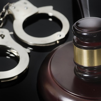
A Los Angeles radiologist has been arrested on suspicion of attempted murder after driving a Tesla sedan with his family inside over a Northern California cliff, according to a report published January 4 by the Daily News.
Dr. Dharmesh Patel, 42, is a radiologist affiliated with Providence Holy Cross Medical Center in Mission Hills, CA. He was arrested on January 2 for attempted murder and child abuse, the Daily News reported from a statement issued on January 3 by the Golden Gate Division of the California Highway Patrol (CHP). Patel will be booked in the San Mateo County jail as soon as he is released from the hospital.
Patel was behind the wheel of the vehicle with his 41-year-old wife, Neha, and their two children, a 7-year-old girl and a 4-year-old boy, on January 2 when the vehicle careened off California's Highway 1 at a spot called Devil's Slide, the Daily News reported. The family's rescue was hindered by a heavy storm and involved helicopters and firefighters rappelling down the cliffside to reach the sedan.
Both children sustained musculoskeletal injuries and were taken to the hospital by ambulance. The adults suffered traumatic injuries and had to be hoisted up the cliff by helicopter; they were then both flown to the hospital, according to the Daily News report.
"After working throughout the night interviewing witnesses and gathering evidence from the scene, authorities said they "have developed probable cause to believe this incident was an intentional act," the CHP said in the statement quoted by the Daily News.


![Overview of the study design. (A) The fully automated deep learning framework was developed to estimate body composition (BC) (defined as subcutaneous adipose tissue [SAT] in liters; visceral adipose tissue [VAT] in liters; skeletal muscle [SM] in liters; SM fat fraction [SMFF] as a percentage; and intramuscular adipose tissue [IMAT] in deciliters) from MRI. The fully automated framework comprised one model (model 1) to quantify different BC measures (SAT, VAT, SM, SMFF, and IMAT) as three-dimensional (3D) measures from whole-body MRI scans. The second model (model 2) was trained to identify standardized anatomic landmarks along the craniocaudal body axis (z coordinate field), which allowed for subdividing the whole-body measures into different subregions typically examined on clinical routine MRI scans (chest, abdomen, and pelvis). (B) BC was quantified from whole-body MRI in over 66,000 individuals from two large population-based cohort studies, the UK Biobank (UKB) (36,317 individuals) and the German National Cohort (NAKO) (30,291 individuals). Bar graphs show age distribution by sex and cohort. BMI = body mass index. (C) After the performance assessment of the fully automated framework, the change in BC measures, distributions, and profiles across age decades were investigated. Age-, sex-, and height-adjusted body composition reference curves were calculated and made publicly available in a web-based z-score calculator (https://circ-ml.github.io).](https://img.auntminnie.com/mindful/smg/workspaces/default/uploads/2026/05/body-comp.XgAjTfPj1W.jpg?auto=format%2Ccompress&fit=crop&h=100&q=70&w=100)


![Overview of the study design. (A) The fully automated deep learning framework was developed to estimate body composition (BC) (defined as subcutaneous adipose tissue [SAT] in liters; visceral adipose tissue [VAT] in liters; skeletal muscle [SM] in liters; SM fat fraction [SMFF] as a percentage; and intramuscular adipose tissue [IMAT] in deciliters) from MRI. The fully automated framework comprised one model (model 1) to quantify different BC measures (SAT, VAT, SM, SMFF, and IMAT) as three-dimensional (3D) measures from whole-body MRI scans. The second model (model 2) was trained to identify standardized anatomic landmarks along the craniocaudal body axis (z coordinate field), which allowed for subdividing the whole-body measures into different subregions typically examined on clinical routine MRI scans (chest, abdomen, and pelvis). (B) BC was quantified from whole-body MRI in over 66,000 individuals from two large population-based cohort studies, the UK Biobank (UKB) (36,317 individuals) and the German National Cohort (NAKO) (30,291 individuals). Bar graphs show age distribution by sex and cohort. BMI = body mass index. (C) After the performance assessment of the fully automated framework, the change in BC measures, distributions, and profiles across age decades were investigated. Age-, sex-, and height-adjusted body composition reference curves were calculated and made publicly available in a web-based z-score calculator (https://circ-ml.github.io).](https://img.auntminnie.com/mindful/smg/workspaces/default/uploads/2026/05/body-comp.XgAjTfPj1W.jpg?auto=format%2Ccompress&fit=crop&h=112&q=70&w=112)


