Digital variance angiography (DVA) can reduce radiation doses to patients versus conventional angiography during certain interventional radiology procedures, a group in Germany has reported.
The finding is from a comparison between the two approaches among patients undergoing treatments for narrowed or blocked arteries in the lower extremities (endovascular peripheral interventions, or EPIs). The technique also improved image quality, noted lead author Till Schürmann, MD, of the University of Freiburg, and colleagues.
"DVA reveals significant radiation dose reduction in lower extremity EPIs and enhances image contrast while decreasing noise," the group wrote. The study was published July 9 in Scientific Reports.
DVA is an emerging motion-based x-ray imaging technique that uses software to visualize the distribution of iodinated contrast medium (ICM) in the vascular system. The technique allows for lower doses of ICM and can be used during standard-of-care angiography examinations, the researchers explained.
To demonstrate its potential advantages, in this study, the group compared its use in 62 patients who underwent EPIs with 370 patients who underwent EPIs with normal ICM dose protocols. They evaluated the overall dose area product (DAP) for each patient from both groups in the pelvic, femoral, and leg regions (popliteal and cruropedal) and assessed image quality by calculating the contrast-to-noise ratio (CNR) in specific regions of interest (ROIs) on vessels.
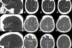
 Pelvic (A), femoral and popliteal (B), and cruro-pedal (C) region. The summated digital variance angiography (DVA) image significantly enhances image quality compared to the digital subtraction angiography (DSA) series I–IV (depicting 4 out of 28 (A), 48 (B) and 28 (C) DSA images) of the low dose (LD) acquisitions. Enhancements of iodinated contrast medium (ICM) by DVA depict small vessel structures in detail while the conventional DSA offers a reduced contrast-to-noise ratio (CNR) using LD acquisitions for standalone diagnostic interpretations.Scientific Reports
According to the analysis, overall DAP was significantly reduced in the DVA group from 3238.6 cGy·cm² to 1230.4 cGy·cm² in pelvic regions, from 1190.9 cGy·cm² to 550.8 cGy·cm² in femoral and popliteal regions, and from 827.6 cGy·cm² to 336.0 cGy·cm² in cruropedal regions.
In addition, DVA images exhibited substantially enhanced ICM contrast and decreased background noise in all regions, the group reported. Specifically, median CNRs in ROIs in the DVA group increased significantly from 8.8 to 14.4 in pelvic regions, from 6.9 to 17.8 in femoral and popliteal regions, and from 7.8 to 17.3 in cruropedal regions.
"DVA can significantly reduce (stochastic) health risk effects of radiation exposure for both patients and staff performing endovascular peripheral interventions," the group wrote.
Ultimately, standard angiography (digital subtraction angiography) remains an essential interventional tool in diagnosing and treating stenosis and occlusions in EPI, and this study adds to the growing evidence of the benefits in DVA, the researchers wrote.
Also, they noted that a limitation of the study was that the image quality evaluation was performed objectively by calculating CNRs, rather than by a subjective qualitative assessment.
"Subjective ratings of the image quality would also be a beneficial determinant for future analysis, especially in prospective studies where categories can be controlled more strictly," the group concluded.
The full study is available here.