A machine-learning model based on chest CT images accurately predicts lung function -- which can then help clinicians better diagnose and assess the severity of chronic obstructive pulmonary disease (COPD).
The findings could improve patient care, as COPD can present in a variety of ways, according to a group of investigators led by doctoral candidate He Sui of China-Japan Union Hospital of Jilin University in Changchun, China. The team's results were published August 14 in the International Journal of Chronic Obstructive Pulmonary Disease.
"Variations in clinical manifestations, physiological characteristics, imaging features, treatment response, disease progression, and survival rates exist among COPD patients, resulting in irreversible pathological changes and permanent damage to the respiratory system," the group wrote. "Therefore, timely diagnosis and early treatment are crucial for delaying disease progression, extending patient lifespan, and improving quality of life."
A pulmonary function test (i.e., spirometry) is the go-to for diagnosing COPD and evaluating its severity, but it may not be able to detect emphysema or airway lesions. It can also be tricky for patients, as some may have difficulty "completing routine pulmonary function tests during the acute stage of COPD or if they have pneumothorax or cardiovascular and cerebrovascular diseases," the authors explained. That's where CT imaging comes in, as it allows for "visual and quantitative evaluation of emphysema presence, pattern, and degree in vivo."
CT postprocessing technology can offer more detailed information about a patient's lung anatomical structure and pathophysiology. For this study, Sui and colleagues developed a machine-learning model that used chest CT images for automated diagnosis and grading of COPD patients. The research included data collected between December 2017 and June 2023 from 173 COPD patients and 176 healthy controls. The investigators used the deep-learning model to segment CT images for lung parenchyma, airway, pulmonary artery, and vein, then extracted imaging features from the segmented regions.
The group found the following:
Performance of machine-learning model for COPD diagnosis and severity grading | ||
Measure | Training set | Test set |
COPD diagnosis | ||
| AUC | 0.98 | 0.97 |
| Accuracy | 95% | 96% |
COPD severity grading | ||
| AUC | 0.89 | 0.8 |
| Accuracy | 78% | 72% |
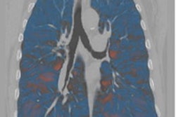
 Image segmentations of lung parenchyma, airway, arteries and veins in COPD patients and healthy controls. Image courtesy of the International Journal of Chronic Obstructive Pulmonary Disease under the Creative Commons Attribution - Noncommercial 4.0 License.
The study results highlight the efficacy of machine learning used with CT imaging to identify and grade COPD, according to the authors, who wrote that, "in comparison with quantitative CT methods reported in previous studies within this field, our developed machine learning model exhibited significantly superior diagnostic performance for COPD patients," and that the findings hold "immense significance in terms of delaying disease progression, extending patient lifespan, and enhancing overall quality of life."
The complete study can be found here.


![Images show the pectoralis muscles of a healthy male individual who never smoked (age, 66 years; height, 178 cm; body mass index [BMI, calculated as weight in kilograms divided by height in meters squared], 28.4; number of cigarette pack-years, 0; forced expiratory volume in 1 second [FEV1], 97.6% predicted; FEV1: forced vital capacity [FVC] ratio, 0.71; pectoralis muscle area [PMA], 59.4 cm2; pectoralis muscle volume [PMV], 764 cm3) and a male individual with a smoking history and chronic obstructive pulmonary disorder (COPD) (age, 66 years; height, 178 cm; BMI, 27.5; number of cigarette pack-years, 43.2, FEV1, 48% predicted; FEV1:FVC, 0.56; PMA, 35 cm2; PMV, 480.8 cm3) from the Canadian Cohort Obstructive Lung Disease (i.e., CanCOLD) study. The CT image is shown in the axial plane. The PMV is automatically extracted using the developed deep learning model and overlayed onto the lungs for visual clarity.](https://img.auntminnie.com/mindful/smg/workspaces/default/uploads/2026/03/genkin.25LqljVF0y.jpg?auto=format%2Ccompress&crop=focalpoint&fit=crop&h=167&q=70&w=250)