Major-minor axis and the mind of the physician
I am reminded of a case that came up when I was an oral examiner for the American Board of Radiology: I don't recall that any clinical information was provided. There was a series of abdominal CT scans (of really poor quality) with a lot of fuzziness in the prevertebral space. Waffling on about lymphomas got zero points, and if a scholar started discoursing on retroperitoneal fibrosis, there was a negative score. The only acceptable answer would be something like the following: "I don't know what this is, but if the patient has any pain or is shocky, I'd really worry about a leaking aortic aneurysm."
 |
| Dr. Jason Birnholz. |
A related issue is the basic, probably universal conditioning of medical education, which is to always be on the lookout for observable signs or a historic indication of an occult systemic process that demands priority in a potential treatment plan. Of course, now everyone tends to rely on labs tests for things such as diabetes, anemia, and causes of acute chest pain and, to an increasing extent, on imaging studies for unsuspected cardiovascular and neoplastic problems.
I tend to think of a major versus minor disease axis. The "cervix" patients have minor disease, and everyone is happy: the patient, because it is not tumor disease and a definite cure for a nagging problem is at hand; the referring physician, because there is an easy fix without further referrals; and, of course, the radiologist in finding something completely unsuspected.
The issue for us is that many physicians limit their referrals to radiology ultrasound facilities to occasions when they are worried about a major risk. The result is that our nice, safe, fast, and exceptionally informative technique is typically underutilized in the community where it can do the most good for individual patient care. There is a myth that a major disease is somehow worth lots of dollars and multiple tests, whereas minor problems (as well as early diagnosis and preventive measures) don't warrant similar expenditures or have an equal chance of reimbursement.
Tiers of care
There is another similar practice gradient, which is to apply most available resources to fixing immediate, local, well-defined, and clinically obvious issues and deferring occult, systemic, and often chronic considerations to a smaller contingent of the system. This is a spectrum from continuous single-provider care for an individual patient to a single visit (such as to an emergency room) focused on a specific issue, such as repair of a laceration.
Medicine is inherently conservative. The first aphorism of Hippocrates says, "Try your best to help, but, above all, do no harm." This is utterly basic. Healthcare systems always need to reconcile this dictum with delivery of services, including instances when inaction can be harmful.
When I was a student at Johns Hopkins there were nurse midwife and nurse anesthetist programs, and during my residency at Massachusetts General Hospital, there was the start of primary care as a "new specialty." These are practical ways to expand the practitioner side of the medical workforce. In essence, this is a trickle down from the educational top with control and monitoring, evolving over the many years into multiple levels of care from nursing, physician's assistants, and self-help resources in medically underserved communities. These have been assuming ever increasing responsibilities in care, typically at the perceived minor disease/immediate limited-care end of these two gradients.
Ultrasound and the 3rd gradient
Diagnostic ultrasound also has a tiered delivery system, but it is unbalanced. Various forms of ultrasound were removed from an imaging science base into different areas of clinical application before the method had stabilized technically. This has served to expand the availability of this service greatly while severely limiting the potential scope of ultrasound as a diagnostic tool.
In many instances, a technologist operates alone, without expert supervision, acquiring images and engaging in interpretation either directly or by selecting particular images to retain or copy for an interpreter to review. The isolated examiner often limits the exam to a specific anatomic region for a study indication perhaps all too often made by a physician, nurse, or office assistant who does not have current understanding of the capabilities of the method. A common example might be "r/o gallstone" rather than "abdominal ultrasound (please) to evaluate right upper quadrant pain."
 |
The third, admittedly arbitrary gradient is between an exploratory "examination," which is in essence a medical consultation, and a simple "test" that is something that might be automated and for which their image acquisition and diagnosis are separable.
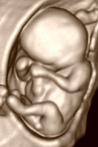
This notion is illustrated by the previous image, which is a 3D image of a 24-mm fetus. Do we evaluate this fetus in its entirety, or do we just seek a nuchal thickness measurement?
Previous page | 1 | 2 | 3 | Next page





![Comparative PET/CT scan images showing [18F]FAPI and [18F]FDG imaging with MIP projections, fusion views, and CT cross-sections for medical diagnosis](https://img.auntminnie.com/mindful/smg/workspaces/default/uploads/2026/10/fapi-vs-fdg.nL6Acp6W2i.jpg?auto=format%2Ccompress&dpr=2&fit=crop&h=167&q=70&w=250)


