Dear AuntMinnie Member,
Attention has been focused on dose reduction for CT exams, but a multicenter team from the U.S. and Switzerland has made an eye-popping achievement: reducing dose for lung follow-up studies to 3% of what's usually used.
The researchers focused on lung exams because patients with lung cancer frequently require multiple studies to follow the course of their disease. They developed a low-dose scanning protocol that reduced dose dramatically without significantly impacting the ability of radiologists to find lung nodules.
Find out how they did it by clicking here, or visit our CT Digital Community at ct.auntminnie.com.
Uncertain future for radiologists?
In other news, radiologists face an uncertain future in a healthcare environment beset by challenges, according to an article by staff writer Kate Madden Yee in our Imaging Leaders Digital Community.
The story profiles a presentation by Dr. Lawrence Muroff at last week's AHRA meeting, in which Dr. Muroff describes what he sees as the eight most challenging trends facing radiologists. These range from declining reimbursement to greater competition from academia.
Find out what impact he thinks these trends will have on radiology by clicking here, or visit our Imaging Leaders Digital Community at leaders.auntminnie.com.
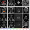


![Examples of ultrasound findings and techniques. (A) Images in a 39-year-old male patient with a mass in the left thigh. The mass is heterogeneous on the B-mode US image (compared with the patient in D) and showed increased microvascularity (superb microvascular imaging [SMI]) and shear-wave elastography (SWE) values. Undifferentiated pleomorphic sarcoma was diagnosed at biopsy (with pleomorphic rhabdomyosarcoma in surgical specimen). (B) Images in an 18-year-old male patient with a mass in the left leg. The mass is hypoechoic on the B-mode image, with no other findings suggestive of malignancy. The lesion is in contact with the cortex of the tibia, which is slightly irregular. CT revealed a doubtful anteromedial tibial erosion. The microvascular study demonstrated high vascularization, suggestive of malignancy. Periosteal Ewing sarcoma was diagnosed with both histologic and immunohistochemical confirmation. (C) Images in a 69-year-old female patient with a lump growing on the outside of the left leg. Multiple SWE examinations were performed (please note the high values obtained in the measurements, whereas the color map highlights the stiffness relative to adjacent tissues). SMI showed areas of increased vascularization to target for sampling. Undifferentiated spindle cell sarcoma was diagnosed at biopsy, with residual leiomyosarcoma in the surgical specimen after neoadjuvant therapy. (D) Images in a 56-year-old female patient with a mass in the right thigh. The mass is heterogeneous at both B-mode ultrasound (similar to patient A) and MRI (coronal T2-weighted spectral attenuated inversion recovery [SPAIR]; T1-weighted pre-contrast and postcontrast imaging), which even shows uptake after the administration of paramagnetic contrast material, which is traditionally suggestive of malignancy. Low values at SMI and elastography are suggestive of benignity. Spindle cell lipoma was diagnosed at biopsy, with atypical spindle cell lipomatous tumor in the surgical specimen.](https://img.auntminnie.com/mindful/smg/workspaces/default/uploads/2026/08/images-radiol250278fig2.APCFLSvX6p.jpg?auto=format%2Ccompress&fit=crop&h=112&q=70&w=112)


