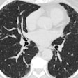
In the study, 27 patients with NSCLC underwent whole-body PET/CT for either primary tumor staging or restaging after neoadjuvant therapy.
The assessment that PET and CT make a dynamic duo for lung cancer staging has been reinforced with the results of a new study. Researchers from University Hospital in Essen, Germany, concluded that dual-modality PET/CT is more accurate than either method alone.
Dr. Gerald Antoch, Dr. Jörg Stattus, Dr. Andre Nemat and colleagues examined 26 patients using both modalities, interpreting the results both separately and in combined images. They found that their accuracy in staging non small-cell lung cancer (NSCLC), which accounts for some 80% of bronchogenic malignancies, increased significantly with the use of a combined scanning device compared to PET and CT by themselves (Radiology, November 2003, Vol. 229:2, pp. 526-533).
CT, MRI, radionuclide scintigraphy and PET have all been used for NSCLC staging. "Although tumor size and infiltration of adjacent structures are accurately assessed with CT, PET has been shown to be substantially more sensitive and specific in the detection and characterization of metastases to mediastinal lymph nodes," the authors wrote. "The limited anatomic information yielded by PET can be overcome by fusing functional PET data with morphologic CT data. Positional and motion-induced data misregistration, however, renders the image fusion of separately acquired CT and PET image sets unsatisfactory."
However, they added, the new dual-modality scanners not only enhance the accuracy of image fusion, they reduce exam times by as much as 30%.
In the study, 27 patients with NSCLC (23 men and 4 women, mean ages 57 and 48, respectively) underwent whole-body PET/CT for either primary tumor staging (n=19) or restaging after neoadjuvant therapy (n=8). Three experienced reader teams evaluated each of the datasets, and all findings were compared to histopathology, the authors wrote.
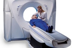
A biograph system (Siemens Medical Solutions, Malvern, PA) was used to perform combined whole-body PET/CT imaging. CT images were acquired from the head to the upper thigh using settings of 130 mAs, 130 kVp, section widths of 5 mm, and table feed of 8 mm rotation (gantry rotation of 0.8 sec.). One hundred forty ml (300 mg/ml) of Iobitridol intravenous contrast (Xenetix, Guerbet, Roissy, France) was administered to all patients; 1.5 g/100 ml of oral barium sulfate contrast was also used in all patients to ensure bowel opacification (Micropaque CT, Guerbet).
Whole-body PET images were acquired an hour after administration of 350 MBq of fluorodeoxyglucose, with patients resting supine during the uptake period to avoid muscular tracer accumulation. Attenuation correction was based on CT data, and PET images were also corrected for scatter and iteratively reconstructed, the authors wrote.
"Tumor staging with CT, PET, and PET/CT was based on the newly revised AJCC TNM system for the classification of lung cancers," the authors wrote. "Cancers of stages IIb are considered resectable, whereas stage IIIb and IV cancers are consisdered nonresectable."
The datasets were analyzed on a workstation (Systrium Technologies, Minneapolis, MN) which provided interactively multiplanar reformations and any desired window settings. Each of 3 reader teams of radiologists and nuclear medicine specialists had the same information about the patients' clinical histories, with the team assessing the combined images consisting of both a radiologist and a nuclear medicine specialist. A McNemar test for matched pairs proportions was used to determine statistical differences in overall AJCC and N stages, with P < .05 considered statistically significant.
According to the T staging results, staging based on PET/CT was more accurate than that based on individual-modality datasets. PET/CT staged 15 patients accurately (with just one understaging). PET correctly staged 12 patients (with 2 overstaged, 2 understaged) and CT also correctly staged 12 patients (with overstaging in 3, and understaging in 1).
As for N staging, PET/CT correctly assessed hilar and mediastinal lymph node involvement in 25/27 patients (overstaging in 1, understaging in 1). CT data alone was correct in 16/27 patients (overstaging in 8, understaging in 3), while PET alone was correct in 24/27 patients (with overstaging in 2, understaging in 1).
"Overall accuracies for the detection of metastasis to lymph nodes were 63% with CT, 89% with PET, and 93% with PET/CT," the authors stated. "Although differences in accuracy between CT and PET/CT (P=.004) and between CT and PET (P=.039) were statistically significant, differences in accuracy between PET and PET/CT were not (P=.625)."
The group found 14 distant metastases in four patients based on CT data, 4 metastases in two patients with PET, and 17 metastases when the modalities were combined.
Overall tumor stage was classified correctly in 19 patients based on CT alone. PET alone proved accurate in 20 patients, and PET/CT led the way with 20 patients staged correctly according to the AJCC tumor stage.
Finally, only PET/CT revealed important incidental findings, including a thrombosis of the right jugular vein in conjunction with central pulmonary artery embolism in one patient, and a local recurrence of rectal carcinoma in a second patient. In a third patient, PET/CT detected hypopharyngeal carcinoma with local lymph node involvement.
Overall, the results confirmed the superiority of PET in the assessment of mediastinal and hilar lymph node metastases, as well as the predominance of CT in the detection of distant metastases, the authors wrote.
"Compared with PET data alone, PET/CT findings led to a change in tumor stage in 7 patients (26%)," they stated. For four patients (15%), this change directly affected the treatment plan. Compared with CT data alone, PET/CT data enabled more accurate assessment of tumor stage in eight patients (30%), which resulted in altered therapeutic recommendations for five patients. "....dual modality PET/CT represents the most accurate approach to NSCLC staging, with a profound effect on therapy, and hence, patient prognosis," the authors concluded.
By Eric BarnesAuntMinnie.com staff writer
November 17, 2003
Related Reading
Financial analysis brings new perspectives on PET/CT acquisition, November 14, 2003
Targeted high-dose radiotherapy benefits inoperable lung cancer patients, November 13, 2003
Lung cancer screening helps smokers kick the habit, October 21, 2003
FLT-PET shows promise in differentiating unclear lung lesions, October 1, 2003
Pairing of CT and PET scans may aid early lung cancer detection, August 22, 2003
PET imaging predicts response to chemotherapy for non-small cell lung cancer, August 4, 2003
Integrated PET-CT better than either alone at staging non-small-cell lung cancer
Copyright © 2003 AuntMinnie.com


