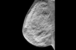
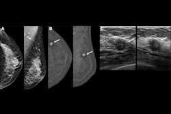
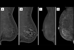
The combination of contrast-enhanced mammography (CEM) and digital breast tomosynthesis (DBT) increases early breast cancer detection each year in women with a personal history of the disease – although it also increases the recall rate, researchers have found.
"Adding contrast-enhanced mammography (CEM) to digital breast tomosynthesis substantially improved the detection of early breast cancer in women with a personal history of breast cancer, and this benefit appears to persist each year," wrote a team led by Wendie Berg, MD, PhD, of the University of Pittsburgh in Pennsylvania in an article published April 30 in Radiology.
The findings are good news for women with a history of breast cancer, as DBT can be insufficient for screening them, Berg's group explained. An ongoing trial called Tomosynthesis or Contrast-Enhanced Mammography (TOCEM) is investigating the use of three annual screenings with both DBT and contrast-enhanced mammography (CEM) for women with breast cancer history; the group conducted a study using data from this trial.
Berg's and colleagues' study tracked three years of results (October 2019 to December 2022), with 1,273 women participating in year one, 819 in year two, and 227 in year three; all of the women underwent a DBT exam and a CEM exam. Two radiologists interpreted both examinations. Berg's group evaluated the effect of adding CEM to DBT on incremental cancer detection rate, cancer type and node status, and recall rate.
The investigators found the following:
| Comparison of DBT alone and DBT plus CEM | ||
|---|---|---|
| Measure | DBT alone | DBT plus CEM |
| Year 1 | ||
| Cancer yield per 1,000 exams | 15.7 | 22.8 |
| Recall rate | 8.1% | 14.7% |
| Year 2 | ||
| Cancer yield per 1,000 exams | 3.7 | 7.3 |
| Recall rate | 3.9% | 9% |
| Year 3 | ||
| Cancer yield per 1,000 exams | 4.4 | 8.8 |
| Recall rate | 3.5% | 7.9% |
The group also reported that 72% of cancers identified were invasive.
Despite the increase in recall rate, adding CEM to DBT shows promise for catching more cancer in women with a personal history of it, according to the authors.
"Our interim results, while preliminary, support the use of CEM for annual supplemental screening in women with a personal history of breast cancer," they concluded.
The complete study can be found here.