Deep learning can help radiologists detect breast cancer on contrast-enhanced CT, according to Japanese research published on October 13 in Clinical Radiology.
A team led by Koichiro Yasaka, PhD, from the University of Tokyo Hospital found that a deep-learning model significantly improved the performance of radiologists interpreting breast CT images.
“Based on these results, it can be inferred that deep learning would be helpful for radiologists and radiology residents working in institutions dealing with patients who have a potentially higher risk for breast cancer, such as older age, genetic mutations, family history of cancer, or smoking,” Yasaka and colleagues wrote.
While CT is not the go-to imaging modality for evaluating breast abnormalities, the researchers pointed out that incidental breast lesions that need further assessment were observed in up to 5.8% of women undergoing chest CT. They added that with the use of CT increasing in daily clinical practice, precise diagnosis of incidental breast cancer on CT could further reduce the associated mortality.
While previous studies suggest deep learning can improve CT detection of incidental breast cancer, the researchers noted that quantitative CT radiomics parameters are affected by intervendor variability.
The Yasaka team investigated whether deep learning could improve the diagnostic performance of radiologists and radiology residents in detecting breast cancer on contrast-enhanced chest CT using data from different vendors in the test stage. It tested its deep-learning model with CT scanners from Canon Medical System and GE HealthCare. It also performed deep learning using preprocessed CT images of the unilateral breast as input data and the binary data for the presence of breast cancer as reference data.
The researchers included patients with confirmed breast cancer who were categorized as the training (n = 201), validation (n = 26), and testing groups (n = 30) using processed CT images from either vendor. They applied the trained deep-learning model to test group patients with breast cancer, which included 30 females with an average age of 59, and patients without breast cancer, which included 19 males and 21 females with an average age of 64 years.
The team found that the model had high performance on both validation and test data. This included area under the curve (AUC) values of 0.976 and 0.967, respectively.
The team also tested the model on the performance of five radiologists with a range of one to 12 years of experience. It found that the model led to significant improvement in the performance of radiologists, represented by a figure of merit values (p = 0.038).
Performance of radiologists with and without deep-learning assistance
| Radiologists | Without deep-learning assistance | With deep-learning assistance |
| Radiologist 1 | 0.933 | 0.958 |
| Radiologist 2 | 0.962 | 0.968 |
| Radiologist 3 | 0.883 | 0.917 |
| Radiologist 4 | 0.944 | 0.947 |
| Radiologist 5 | 0.867 | 0.900 |
The study authors also reported that the radiologists believed that the deep-learning model was “more useful” in diagnosing patients with breast cancer than diagnosing patients with lesions apart from breast cancer.
They also called for larger prospective studies to further validate their model. This includes studies using CT scanners from more than two vendors.
Still, they highlighted that their results show deep learning’s promise for helping radiologists and radiology residents in finding breast cancer on contrast-enhanced chest CT.
The full study can be found here.

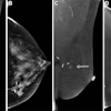

![A normal mammogram confirmed by three-year radiologic follow-up illustrates reader-marked regions of interest (ROIs) during (A) unaided (round 1) and (B) artificial intelligence (AI)–assisted (round 2) reading. Each colored dot represents an ROI for recall by a human reader. Readers could mark more than one ROI per case, represented by multiple dots of the same color. During AI-assisted reading, the AI system displayed three visible prompts: two with suspicion of malignancy scores of 35% (left mediolateral oblique [L MLO] and craniocaudal [L CC]) and one with a suspicion of malignancy score of 10% (right craniocaudal [R CC]), shown as polygonal overlays. Without AI, six of 10 readers (60%) marked a false-positive ROI. With AI assistance, this fell to two of 10 (20%). R MLO = right mediolateral oblique.](https://img.auntminnie.com/mindful/smg/workspaces/default/uploads/2026/07/2026-07-14-radiology-mammogram-ai-auto-bias.H0bYO8QlWs.jpg?auto=format%2Ccompress&fit=crop&h=100&q=70&w=100)


![A normal mammogram confirmed by three-year radiologic follow-up illustrates reader-marked regions of interest (ROIs) during (A) unaided (round 1) and (B) artificial intelligence (AI)–assisted (round 2) reading. Each colored dot represents an ROI for recall by a human reader. Readers could mark more than one ROI per case, represented by multiple dots of the same color. During AI-assisted reading, the AI system displayed three visible prompts: two with suspicion of malignancy scores of 35% (left mediolateral oblique [L MLO] and craniocaudal [L CC]) and one with a suspicion of malignancy score of 10% (right craniocaudal [R CC]), shown as polygonal overlays. Without AI, six of 10 readers (60%) marked a false-positive ROI. With AI assistance, this fell to two of 10 (20%). R MLO = right mediolateral oblique.](https://img.auntminnie.com/mindful/smg/workspaces/default/uploads/2026/07/2026-07-14-radiology-mammogram-ai-auto-bias.H0bYO8QlWs.jpg?auto=format%2Ccompress&dpr=2&fit=crop&h=167&q=70&w=250)


