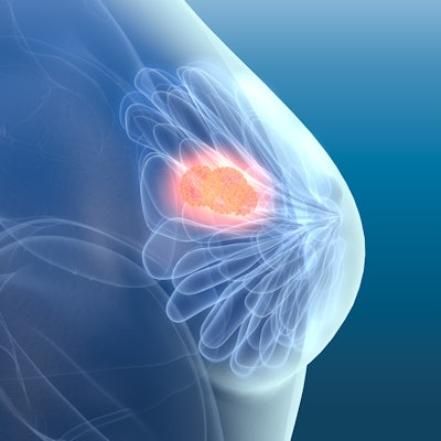
QT Imaging is highlighting a study that used its QTscan technology to analyze changes in breast glandular and ductal volume during the menstrual cycle in premenopausal, perimenopausal, and postmenopausal women.
Study collaborators at the Dr. Susan Love Research Foundation analyzed over 400 images of 24 women acquired with the company's QT Imaging breast scanner. The women had no identified breast malignancies during their menstrual cycle.
Results showed that glandular tissue has greater menstrual cycle variation in premenopausal breasts than in postmenopausal breasts, whereas ductal tissue does not have significantly more variation in premenopausal breasts than in postmenopausal breasts, according to the company.
The study was presented at the SPIE Medical Imaging Conference in February in San Diego and provides a more accurate description of tissue changes during the menstrual cycle. Importantly, results showed quantitatively different behavior in glandular and ductal tissue in vivo, which is the first time such an observation has been made, QT Imaging said.
The QT Imaging Breast Scanner is indicated for use as an ultrasonic imaging system to provide reflection-mode and transmission-mode images of a patient's breast.

![A normal mammogram confirmed by three-year radiologic follow-up illustrates reader-marked regions of interest (ROIs) during (A) unaided (round 1) and (B) artificial intelligence (AI)–assisted (round 2) reading. Each colored dot represents an ROI for recall by a human reader. Readers could mark more than one ROI per case, represented by multiple dots of the same color. During AI-assisted reading, the AI system displayed three visible prompts: two with suspicion of malignancy scores of 35% (left mediolateral oblique [L MLO] and craniocaudal [L CC]) and one with a suspicion of malignancy score of 10% (right craniocaudal [R CC]), shown as polygonal overlays. Without AI, six of 10 readers (60%) marked a false-positive ROI. With AI assistance, this fell to two of 10 (20%). R MLO = right mediolateral oblique.](https://img.auntminnie.com/mindful/smg/workspaces/default/uploads/2026/07/2026-07-14-radiology-mammogram-ai-auto-bias.H0bYO8QlWs.jpg?auto=format%2Ccompress&fit=crop&h=100&q=70&w=100)


![A normal mammogram confirmed by three-year radiologic follow-up illustrates reader-marked regions of interest (ROIs) during (A) unaided (round 1) and (B) artificial intelligence (AI)–assisted (round 2) reading. Each colored dot represents an ROI for recall by a human reader. Readers could mark more than one ROI per case, represented by multiple dots of the same color. During AI-assisted reading, the AI system displayed three visible prompts: two with suspicion of malignancy scores of 35% (left mediolateral oblique [L MLO] and craniocaudal [L CC]) and one with a suspicion of malignancy score of 10% (right craniocaudal [R CC]), shown as polygonal overlays. Without AI, six of 10 readers (60%) marked a false-positive ROI. With AI assistance, this fell to two of 10 (20%). R MLO = right mediolateral oblique.](https://img.auntminnie.com/mindful/smg/workspaces/default/uploads/2026/07/2026-07-14-radiology-mammogram-ai-auto-bias.H0bYO8QlWs.jpg?auto=format%2Ccompress&dpr=2&fit=crop&h=167&q=70&w=250)


