Digital breast tomosynthesis (DBT) is best known for its ability to detect breast cancer, but could the modality also assess bone health? A December 13 study in Bone described how a Michigan research team used DBT to perform bone density measurement.
The method described in the study could make osteoporosis screening more widely available. While many guidelines recommend bone imaging for women over the age of 65 with a major osteoporosis risk factor, only about one-third of women undergo osteoporosis screening after an initial low-energy break.
In contrast, upward of 90% of women in this age group follow recommended screening mammography schedules. The authors posited that the osteoporosis screening rate could be boosted by offering bone assessment at the same time --- and with the same imaging modality -- as breast screening.
"Digital wrist tomosynthesis is feasible in a mammography setting, and informative on bone mass, cortical thickness, and microstructural qualities that are known to deteriorate in osteoporosis," wrote the authors, led by Yener Yeni, PhD, a biomechanics researcher at the Henry Ford Health System Bone and Joint Center in Detroit. "To our knowledge, this study represents the first application of DBT for imaging bone."
Yeni and colleagues first tested the feasibility of using DBT for bone imaging with five cadaver forearms. They scanned the arms using a DBT system, dual-energy x-ray absorptiometry (DEXA), and micro-CT in order to establish baseline measurements across modalities.
After confirming ex-vivo feasibility, the team recruited five patients, ranging in age from 19 to 75, to undergo wrist tomosynthesis scans. To acquire the digital wrist tomosynthesis (DWT) images, the patients aligned their nondominant, left hand on a generic hand template the researchers taped to a DBT machine.
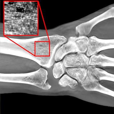
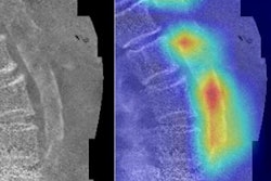
 Left, a 19-year-old woman undergoing a wrist imaging using a DBT scanner. Right, DWT image of same patient. Image courtesy of Yener Yeni, PhD.
The team acquired DWT images three times, repositioning the patients' arms to acquire views from different angles. The researchers then used computer calculations to quantify bone mineral density metrics from the DWT images.
The tomosynthesis bone measurements strongly correlated with those from DEXA and micro-CT scans. Even with the small sample size, metrics such as bone mass, cortical thickness, and integral bone volume fraction all correlated with the reference measurements on a statistically significant basis.
Furthermore, the DWT metrics achieved sufficient repeatability, and the majority of structures hit the 0.7 threshold needed to demonstrate a strong positive correlation with reference metrics.
The DWT metric of bone volume fraction, in particular, looked like a useful correlate for bone mineral density. Bone volume fraction was not only significantly associated with patient age in the study but also was similar to volumetric measurements on CT and bone mineral density measurements on DEXA.
Equally important, the researchers found the DWT image acquisition process was suitable for adding to a mammography appointment. Neither the patients nor staff reported difficulty with the imaging protocol, and a dosimeter showed a radiation dose comparable to that of a standard radiographic exam. The session also took less than five minutes from entrance and exit.
The findings demonstrate the promise of using a DBT system to assess bone health among mammography patients, but more research is needed before DWT goes mainstream. Researchers still need to calibrate DWT measurements to those derived from DEXA scans, and the authors called for more powerful, case-control studies.
Should researchers figure out the technicalities, digital wrist tomosynthesis could benefit numerous mammography patients, including obese women, for whom traditional scans aren't as effective, and breast cancer survivors, who are at increased risk of bone loss. The best part? The technology is widely available, and patients are already coming in for mammograms.
"It is reasonable to expect that with increasing adoption of DBT and continued high adherence to breast screening, bone screening would become more prevalent if offered in coordination with DBT breast exams, provided that osteoporosis can be assessed using a DBT modality," the authors wrote.
![A normal mammogram confirmed by three-year radiologic follow-up illustrates reader-marked regions of interest (ROIs) during (A) unaided (round 1) and (B) artificial intelligence (AI)–assisted (round 2) reading. Each colored dot represents an ROI for recall by a human reader. Readers could mark more than one ROI per case, represented by multiple dots of the same color. During AI-assisted reading, the AI system displayed three visible prompts: two with suspicion of malignancy scores of 35% (left mediolateral oblique [L MLO] and craniocaudal [L CC]) and one with a suspicion of malignancy score of 10% (right craniocaudal [R CC]), shown as polygonal overlays. Without AI, six of 10 readers (60%) marked a false-positive ROI. With AI assistance, this fell to two of 10 (20%). R MLO = right mediolateral oblique.](https://img.auntminnie.com/mindful/smg/workspaces/default/uploads/2026/07/2026-07-14-radiology-mammogram-ai-auto-bias.H0bYO8QlWs.jpg?auto=format%2Ccompress&fit=crop&h=100&q=70&w=100)


![A normal mammogram confirmed by three-year radiologic follow-up illustrates reader-marked regions of interest (ROIs) during (A) unaided (round 1) and (B) artificial intelligence (AI)–assisted (round 2) reading. Each colored dot represents an ROI for recall by a human reader. Readers could mark more than one ROI per case, represented by multiple dots of the same color. During AI-assisted reading, the AI system displayed three visible prompts: two with suspicion of malignancy scores of 35% (left mediolateral oblique [L MLO] and craniocaudal [L CC]) and one with a suspicion of malignancy score of 10% (right craniocaudal [R CC]), shown as polygonal overlays. Without AI, six of 10 readers (60%) marked a false-positive ROI. With AI assistance, this fell to two of 10 (20%). R MLO = right mediolateral oblique.](https://img.auntminnie.com/mindful/smg/workspaces/default/uploads/2026/07/2026-07-14-radiology-mammogram-ai-auto-bias.H0bYO8QlWs.jpg?auto=format%2Ccompress&dpr=2&fit=crop&h=167&q=70&w=250)


