GE Healthcare and St. Luke's University Health Network in Pennsylvania will pilot the first One-Stop Clinic rapid diagnostic breast cancer center model in the U.S.
The One-Stop Clinic model utilizes a multimodality approach to coordinate patient journeys from initial appointment through diagnosis and treatment plan. It keeps patients with one team in one location to reduce the time frame between an abnormal screening mammogram and a breast cancer treatment plan.
The Gustave Roussy Cancer Center in France first created the model to improve clinical outcomes and expedite breast cancer diagnosis and treatment planning. GE Healthcare and Premier previously announced plans in 2019 to open the first One-Stop Clinic model in the U.S.
GE Healthcare and Premiere Applied Sciences chose St. Luke's for the pilot site because the health network already employs a five-day diagnostic imaging workflow. Once operational, the One-Stop Clinic will further reduce this time to 36 hours or fewer.
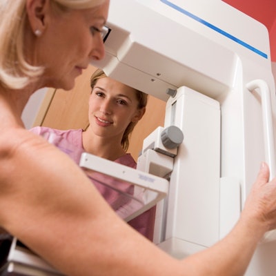
St. Luke's has begun to pilot the shorter workflow and plans to open the One-Stop Clinic in 2021. The patient's journey through the clinic will start with a mammogram, followed by a biopsy and pathology as needed, with diagnosis and treatment planning services to women with confirmed cancer cases.
The goal is to take what's learned from St. Luke's and share key findings with other health systems in the future. A shorter diagnostic process could also help mitigate breast cancer diagnostic delays due to thousands of missed mammograms during the COVID-19 pandemic.

![A normal mammogram confirmed by three-year radiologic follow-up illustrates reader-marked regions of interest (ROIs) during (A) unaided (round 1) and (B) artificial intelligence (AI)–assisted (round 2) reading. Each colored dot represents an ROI for recall by a human reader. Readers could mark more than one ROI per case, represented by multiple dots of the same color. During AI-assisted reading, the AI system displayed three visible prompts: two with suspicion of malignancy scores of 35% (left mediolateral oblique [L MLO] and craniocaudal [L CC]) and one with a suspicion of malignancy score of 10% (right craniocaudal [R CC]), shown as polygonal overlays. Without AI, six of 10 readers (60%) marked a false-positive ROI. With AI assistance, this fell to two of 10 (20%). R MLO = right mediolateral oblique.](https://img.auntminnie.com/mindful/smg/workspaces/default/uploads/2026/07/2026-07-14-radiology-mammogram-ai-auto-bias.H0bYO8QlWs.jpg?auto=format%2Ccompress&fit=crop&h=100&q=70&w=100)


![A normal mammogram confirmed by three-year radiologic follow-up illustrates reader-marked regions of interest (ROIs) during (A) unaided (round 1) and (B) artificial intelligence (AI)–assisted (round 2) reading. Each colored dot represents an ROI for recall by a human reader. Readers could mark more than one ROI per case, represented by multiple dots of the same color. During AI-assisted reading, the AI system displayed three visible prompts: two with suspicion of malignancy scores of 35% (left mediolateral oblique [L MLO] and craniocaudal [L CC]) and one with a suspicion of malignancy score of 10% (right craniocaudal [R CC]), shown as polygonal overlays. Without AI, six of 10 readers (60%) marked a false-positive ROI. With AI assistance, this fell to two of 10 (20%). R MLO = right mediolateral oblique.](https://img.auntminnie.com/mindful/smg/workspaces/default/uploads/2026/07/2026-07-14-radiology-mammogram-ai-auto-bias.H0bYO8QlWs.jpg?auto=format%2Ccompress&fit=crop&h=112&q=70&w=112)