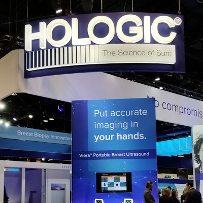
Women's imaging vendor Hologic has issued a statement reaffirming the clinical benefits of digital breast tomosynthesis (DBT) for breast cancer screening in response to a recent USA Today article questioning the exam's value.
Hologic's statement aims to address potential confusion among the public concerning the merits of DBT, or 3D mammography, compared with conventional 2D mammography, in light of allegations made in the news article.
In the story, investigators from Kaiser Health News suggested that mammography vendors, hospitals, physicians, and patient advocates have been promoting the use of DBT -- a more expensive exam than conventional 2D mammography -- for breast cancer screening despite insufficient support from large-scale clinical trials.
Countering these claims, Hologic asserted that more than 250 peer-reviewed clinical studies have demonstrated the diagnostic advantages of DBT over 2D mammography, including the exam's capacity to detect between 20% and 65% more invasive cancers and reduce unnecessary callbacks by as much as 40%.
"3D mammography is unquestionably a superior mammogram," Dr. Nila Alsheik of Advocate Lutheran General Hospital in Park Ridge, IL, said in a statement from Hologic. "In my practice, I still see 2 to 3 centimeter cancers that are completely occult on 2D mammography, and only visualized on 3D mammography."
"The clinical benefits of DBT are indisputable ... [and DBT] is approved as superior for women with dense breasts compared to 2D alone," Hologic said.



![A normal mammogram confirmed by three-year radiologic follow-up illustrates reader-marked regions of interest (ROIs) during (A) unaided (round 1) and (B) artificial intelligence (AI)–assisted (round 2) reading. Each colored dot represents an ROI for recall by a human reader. Readers could mark more than one ROI per case, represented by multiple dots of the same color. During AI-assisted reading, the AI system displayed three visible prompts: two with suspicion of malignancy scores of 35% (left mediolateral oblique [L MLO] and craniocaudal [L CC]) and one with a suspicion of malignancy score of 10% (right craniocaudal [R CC]), shown as polygonal overlays. Without AI, six of 10 readers (60%) marked a false-positive ROI. With AI assistance, this fell to two of 10 (20%). R MLO = right mediolateral oblique.](https://img.auntminnie.com/mindful/smg/workspaces/default/uploads/2026/07/2026-07-14-radiology-mammogram-ai-auto-bias.H0bYO8QlWs.jpg?auto=format%2Ccompress&fit=crop&h=100&q=70&w=100)


![A normal mammogram confirmed by three-year radiologic follow-up illustrates reader-marked regions of interest (ROIs) during (A) unaided (round 1) and (B) artificial intelligence (AI)–assisted (round 2) reading. Each colored dot represents an ROI for recall by a human reader. Readers could mark more than one ROI per case, represented by multiple dots of the same color. During AI-assisted reading, the AI system displayed three visible prompts: two with suspicion of malignancy scores of 35% (left mediolateral oblique [L MLO] and craniocaudal [L CC]) and one with a suspicion of malignancy score of 10% (right craniocaudal [R CC]), shown as polygonal overlays. Without AI, six of 10 readers (60%) marked a false-positive ROI. With AI assistance, this fell to two of 10 (20%). R MLO = right mediolateral oblique.](https://img.auntminnie.com/mindful/smg/workspaces/default/uploads/2026/07/2026-07-14-radiology-mammogram-ai-auto-bias.H0bYO8QlWs.jpg?auto=format%2Ccompress&fit=crop&h=112&q=70&w=112)