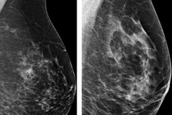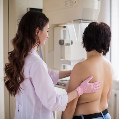
Young women at high risk of breast cancer due to a family or personal history of the disease or having dense breasts benefit from regular mammography screening starting at age 30, according to a study published online September 18 in the Journal of the American College of Radiology.
A team led by Dr. Cindy Lee of the University of California, San Francisco (UCSF) found that women ages 30 to 39 who had these risk factors experienced significantly higher cancer detection rates.
"Our results confirm the positive association of the presence of any of these three risk factors with significantly higher cancer detection rates in women beginning at age 30," the group wrote.
Lee and colleagues' study compared risk-based screening of women ages 30 to 39 with risk factors versus women ages 40 to 49 without risk factors. They used data from 150 National Mammography Database facilities in 31 states for 2.6 million women. The women had undergone screening mammography between January 2008 and December 2015.
The researchers stratified patients by five-year age intervals, availability of prior mammograms, and the following risk factors for breast cancer: family history of breast cancer, personal history of breast cancer, and dense breasts.
They then calculated the recall rate, cancer detection rates, and positive predictive values for biopsy recommended (PPV2) and biopsy performed (PPV3) for each age and risk group.
Lee and colleagues found the following:
- The overall mean cancer detection rate was 3.69 of 1,000.
- The overall mean recall rate was 10%.
- The overall PPV2 was 20.1%.
- The overall PPV3 was 28.2%.
Women ages 30 to 39 had similar cancer detection and recall rates, as well as similar PPVs, with the presence of the three evaluated risk factors associated with significantly higher cancer detection rates.
"Our findings suggest that recommendations of screening mammography should be personalized on the basis of a woman's age, breast density, personal history of breast cancer, and family history of breast cancer," the group concluded. "Specifically, women with any of these three specific risk factors can benefit by starting screening at age 30, instead of at age 40 as currently considered for women without risk factors."