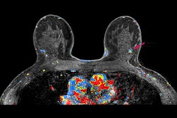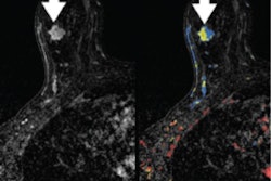
Contrast-enhanced digital mammography (CEDM) performs comparably to or better than digital mammography when it comes to breast cancer screening in women at higher risk of breast cancer, according to a study published online August 27 in Radiology.
The findings suggest that CEDM is an effective additional tool in the breast screening arsenal, wrote a team led by Dr. Janice Sung of Memorial Sloan-Kettering Cancer Center in New York City.
"CEDM has been shown to be more sensitive and specific than two-dimensional full-field digital mammography in the diagnostic setting ... [but] few studies have reported on its performance in the screening setting," the group wrote. "CEDM is a promising technique for screening women with higher-than-average risk for breast cancer."
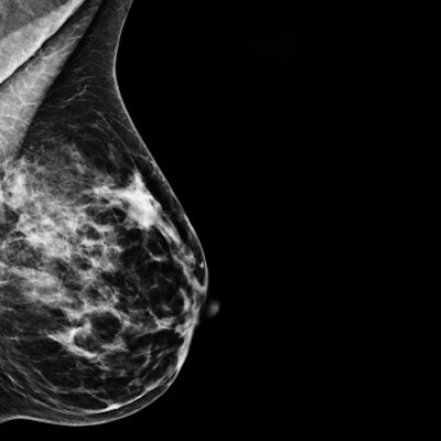
Although mammography is the primary imaging modality for breast cancer screening, it has limitations, specifically in women with dense breast tissue. Breast MRI is known to be the most sensitive modality for finding breast cancer across all tissue densities, but it can be expensive, which makes contrast-enhanced digital mammography an appealing alternative. CEDM has also been shown to perform comparably to breast MRI for detecting cancers not found on conventional mammography, the team noted.
Sung's group evaluated the performance of CEDM compared with digital mammography in a study that included 904 screening CEDM exams performed between December 2012 and April 2016. Of these 904 women, 77.4% had dense breast tissue, 27.3% had a family history of breast cancer, and 40.2% had a personal history of breast cancer.
The team reviewed women's medical records for age, risk factors, short-interval follow-up and biopsies recommended, and cancers detected. They also tracked sensitivity, specificity, positive predictive value of abnormal findings at screening (PPV1), positive predictive value of biopsy performed, and negative predictive value (NPV).
The researchers found that both CEDM's sensitivity and its negative predictive value for screening women at increased risk for breast cancer were higher than those for digital mammography.
| CEDM vs. digital mammography for breast screening | |||
| Performance measure | Digital | CEDM | p-value |
| Sensitivity | 50% | 87.5% | 0.03 |
| Specificity | 97.1% | 93.7% | < 0.001 |
| PPV1 | 25% | 20.9% | 0.39 |
| NPV | 99% | 99.7% | 0.02 |
CEDM's cancer detection rate was 15.5 per 1,000 women. Its positive predictive value of biopsy performed was 29.4%, comparable to benchmarks for both mammography (20% to 45%) and breast MRI (20% to 50%), the group noted.
Screening mammography's limitations have led to a search for improved screening techniques, and CEDM may be just the ticket, the group wrote.
"Our results suggest that CEDM has the potential to be an alternative screening technique to two-dimensional full-field digital mammography in women at increased risk of breast cancer," Sung and colleagues concluded.