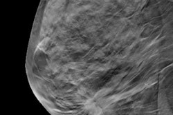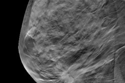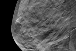
VIENNA - A large study out of Norway presented at ECR 2018 demonstrated that the rate of screen-detected breast cancer was about 50% higher for women screened with synthesized 2D mammography and digital breast tomosynthesis (DBT) compared with digital mammography alone.
The researchers looked at nearly 100,000 women in a population-based screening trial to determine how DBT plus synthesized 2D mammography compares with standard digital mammography alone. Tomosynthesis data are used to reconstruct a 2D mammogram, which is called synthetic 2D mammography.
"We found an increased detection of both [ductal carcinoma in situ (DCIS)] and invasive cancer, including an increased rate of tubular carcinomas," said lead author Dr. Tone Hovda from Drammen Hospital in Drammen, Norway. "The recall rate did not differ."
She and her colleagues also found smaller tumors and lower-grade tumors in the DBT group.
The researchers included women ages 50 to 69 who were screened biennially. The DBT plus synthetic 2D arm encompassed 37,185 women. The standard mammography arm included 61,743 women hailing from screening centers in Vestfold and VestreViken.
| 2D mammo vs. DBT + 2D mammo in Norway | ||
| 2D mammography | DBT + synthetic 2D mammo | |
| Recall rate* | 33 | 33.7 |
| Invasive carcinoma rate | 5.3 | 7.6 |
| DCIS rate | 0.8 | 1.7 |
| Total detection rate | 6.1 | 9.4 |
The researchers also found eight times more tubular carcinomas in the DBT group, although the numbers were small. More grade 1 cancers also showed up in the DBT group versus the standard 2D mammography group.
In terms of future research, Hovda and colleagues are turning their attention toward studying mammographic features, breast density, and interval cancers.

![A normal mammogram confirmed by three-year radiologic follow-up illustrates reader-marked regions of interest (ROIs) during (A) unaided (round 1) and (B) artificial intelligence (AI)–assisted (round 2) reading. Each colored dot represents an ROI for recall by a human reader. Readers could mark more than one ROI per case, represented by multiple dots of the same color. During AI-assisted reading, the AI system displayed three visible prompts: two with suspicion of malignancy scores of 35% (left mediolateral oblique [L MLO] and craniocaudal [L CC]) and one with a suspicion of malignancy score of 10% (right craniocaudal [R CC]), shown as polygonal overlays. Without AI, six of 10 readers (60%) marked a false-positive ROI. With AI assistance, this fell to two of 10 (20%). R MLO = right mediolateral oblique.](https://img.auntminnie.com/mindful/smg/workspaces/default/uploads/2026/07/2026-07-14-radiology-mammogram-ai-auto-bias.H0bYO8QlWs.jpg?auto=format%2Ccompress&fit=crop&h=100&q=70&w=100)


![A normal mammogram confirmed by three-year radiologic follow-up illustrates reader-marked regions of interest (ROIs) during (A) unaided (round 1) and (B) artificial intelligence (AI)–assisted (round 2) reading. Each colored dot represents an ROI for recall by a human reader. Readers could mark more than one ROI per case, represented by multiple dots of the same color. During AI-assisted reading, the AI system displayed three visible prompts: two with suspicion of malignancy scores of 35% (left mediolateral oblique [L MLO] and craniocaudal [L CC]) and one with a suspicion of malignancy score of 10% (right craniocaudal [R CC]), shown as polygonal overlays. Without AI, six of 10 readers (60%) marked a false-positive ROI. With AI assistance, this fell to two of 10 (20%). R MLO = right mediolateral oblique.](https://img.auntminnie.com/mindful/smg/workspaces/default/uploads/2026/07/2026-07-14-radiology-mammogram-ai-auto-bias.H0bYO8QlWs.jpg?auto=format%2Ccompress&fit=crop&h=112&q=70&w=112)