Friday, December 4 | 11:30 a.m.-11:40 a.m. | SST01-07 | Room E450B
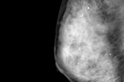
In this Friday session, Dutch investigators will present an automated way to identify women who most need supplemental breast cancer screening, based on breast density.The sensitivity of mammograms is low for women with dense breasts, since cancers may be masked by dense tissue, reported the study team from Radboud University in Nijmegen.
The study examined methods for identifying women with density patterns associated with a high risk of masked breast cancers. The group used three methods based on quantitative volumetric breast density analysis, and compared each one to an area-based density measurement.
"We use large screening databases including over 200,000 exams to investigate methods to identify women with breast density patterns that are likely to mask cancers in mammograms when they develop," explained lead author Katharina Holland in an email to AuntMinnie.com.
These are the women who would benefit most from supplemental screening with ultrasound or MRI, she said.
"We found that existing breast density measurements are able to differentiate between mammograms taken prior to interval cancers and mammograms of normal controls," Holland said. "However, some novel masking measures had a significantly higher performance in identifying women at risk for a masked cancer."
The idea is to implement supplemental screening as an adjunct to mammography with the goal of reducing the number of interval cancers, she added.
"The aim should be to stratify women with a measure that optimally predicts masking potential, while the number of women offered additional imaging remains low," she said. "Our masking measures are a promising step in this direction."

![A normal mammogram confirmed by three-year radiologic follow-up illustrates reader-marked regions of interest (ROIs) during (A) unaided (round 1) and (B) artificial intelligence (AI)–assisted (round 2) reading. Each colored dot represents an ROI for recall by a human reader. Readers could mark more than one ROI per case, represented by multiple dots of the same color. During AI-assisted reading, the AI system displayed three visible prompts: two with suspicion of malignancy scores of 35% (left mediolateral oblique [L MLO] and craniocaudal [L CC]) and one with a suspicion of malignancy score of 10% (right craniocaudal [R CC]), shown as polygonal overlays. Without AI, six of 10 readers (60%) marked a false-positive ROI. With AI assistance, this fell to two of 10 (20%). R MLO = right mediolateral oblique.](https://img.auntminnie.com/mindful/smg/workspaces/default/uploads/2026/07/2026-07-14-radiology-mammogram-ai-auto-bias.H0bYO8QlWs.jpg?auto=format%2Ccompress&fit=crop&h=100&q=70&w=100)


![A normal mammogram confirmed by three-year radiologic follow-up illustrates reader-marked regions of interest (ROIs) during (A) unaided (round 1) and (B) artificial intelligence (AI)–assisted (round 2) reading. Each colored dot represents an ROI for recall by a human reader. Readers could mark more than one ROI per case, represented by multiple dots of the same color. During AI-assisted reading, the AI system displayed three visible prompts: two with suspicion of malignancy scores of 35% (left mediolateral oblique [L MLO] and craniocaudal [L CC]) and one with a suspicion of malignancy score of 10% (right craniocaudal [R CC]), shown as polygonal overlays. Without AI, six of 10 readers (60%) marked a false-positive ROI. With AI assistance, this fell to two of 10 (20%). R MLO = right mediolateral oblique.](https://img.auntminnie.com/mindful/smg/workspaces/default/uploads/2026/07/2026-07-14-radiology-mammogram-ai-auto-bias.H0bYO8QlWs.jpg?auto=format%2Ccompress&fit=crop&h=112&q=70&w=112)