Tuesday, December 1 | 10:10 a.m.-10:20 a.m. | RC315-09 | Arie Crown Theater
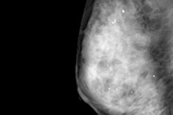
Using synthesized 2D mammography images with digital breast tomosynthesis (DBT) yields recall rates comparable to those of conventional mammography in combination with DBT, researchers from the University of Pennsylvania Health System report.Dr. Samantha Zuckerman and colleagues compared recall rates generated with synthetic 2D plus DBT and conventional mammography plus DBT, and analyzed the types of lesions that were recalled. Between October 2011 and February 2013, 15,571 women were screened with the digital mammography-DBT combination. Meanwhile, 2,090 women were screened with the synthetic 2D-DBT combination between January and March 2015.
The overall recall rate for synthetic 2D-DBT was 8.3%, compared with mammography-DBT's rate of 8.8%, Zuckerman's group found. The two combinations were also comparable in terms of the type of lesion recalled.
The study results show stable recall rates and lesion types between the two combinations, which suggests that replacing conventional 2D mammography with synthesized 2D images maintains recall rates -- while also adding the benefit of reducing radiation dose, Zuckerman's team found.


![A normal mammogram confirmed by three-year radiologic follow-up illustrates reader-marked regions of interest (ROIs) during (A) unaided (round 1) and (B) artificial intelligence (AI)–assisted (round 2) reading. Each colored dot represents an ROI for recall by a human reader. Readers could mark more than one ROI per case, represented by multiple dots of the same color. During AI-assisted reading, the AI system displayed three visible prompts: two with suspicion of malignancy scores of 35% (left mediolateral oblique [L MLO] and craniocaudal [L CC]) and one with a suspicion of malignancy score of 10% (right craniocaudal [R CC]), shown as polygonal overlays. Without AI, six of 10 readers (60%) marked a false-positive ROI. With AI assistance, this fell to two of 10 (20%). R MLO = right mediolateral oblique.](https://img.auntminnie.com/mindful/smg/workspaces/default/uploads/2026/07/2026-07-14-radiology-mammogram-ai-auto-bias.H0bYO8QlWs.jpg?auto=format%2Ccompress&fit=crop&h=100&q=70&w=100)


![A normal mammogram confirmed by three-year radiologic follow-up illustrates reader-marked regions of interest (ROIs) during (A) unaided (round 1) and (B) artificial intelligence (AI)–assisted (round 2) reading. Each colored dot represents an ROI for recall by a human reader. Readers could mark more than one ROI per case, represented by multiple dots of the same color. During AI-assisted reading, the AI system displayed three visible prompts: two with suspicion of malignancy scores of 35% (left mediolateral oblique [L MLO] and craniocaudal [L CC]) and one with a suspicion of malignancy score of 10% (right craniocaudal [R CC]), shown as polygonal overlays. Without AI, six of 10 readers (60%) marked a false-positive ROI. With AI assistance, this fell to two of 10 (20%). R MLO = right mediolateral oblique.](https://img.auntminnie.com/mindful/smg/workspaces/default/uploads/2026/07/2026-07-14-radiology-mammogram-ai-auto-bias.H0bYO8QlWs.jpg?auto=format%2Ccompress&fit=crop&h=112&q=70&w=112)