Monday, November 30 | 12:45 p.m.-1:15 p.m. | BR236-SD-MOB4 | Lakeside Learning Center, Station 4
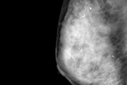
Using reconstructed 2D images with digital breast tomosynthesis decreases radiation dose to patients by more than 40%, according to this Monday afternoon poster presentation.Bruno Barufaldi and colleagues from São Carlos, Brazil, and the University of Pennsylvania examined the impact of using 2D reconstructed mammograms with digital breast tomosynthesis, rather than a combination mode of the two technologies. For the study, the group included 991 women who received either a combination of 2D and 3D mammography or 3D mammography with 2D image reconstruction over two 13-day periods in 2014 and 2015. The researchers calculated the average glandular dose (AGD) using customized software.
During the 2014 time period, 485 combination studies were performed, with an average AGD per view of 4.28 mGy. During the 2015 time period, 506 tomosynthesis exams with 2D image reconstruction were conducted, with an average AGD of 2.42 mGy -- a dose reduction of 43%, according to the group.


![A normal mammogram confirmed by three-year radiologic follow-up illustrates reader-marked regions of interest (ROIs) during (A) unaided (round 1) and (B) artificial intelligence (AI)–assisted (round 2) reading. Each colored dot represents an ROI for recall by a human reader. Readers could mark more than one ROI per case, represented by multiple dots of the same color. During AI-assisted reading, the AI system displayed three visible prompts: two with suspicion of malignancy scores of 35% (left mediolateral oblique [L MLO] and craniocaudal [L CC]) and one with a suspicion of malignancy score of 10% (right craniocaudal [R CC]), shown as polygonal overlays. Without AI, six of 10 readers (60%) marked a false-positive ROI. With AI assistance, this fell to two of 10 (20%). R MLO = right mediolateral oblique.](https://img.auntminnie.com/mindful/smg/workspaces/default/uploads/2026/07/2026-07-14-radiology-mammogram-ai-auto-bias.H0bYO8QlWs.jpg?auto=format%2Ccompress&fit=crop&h=100&q=70&w=100)


![A normal mammogram confirmed by three-year radiologic follow-up illustrates reader-marked regions of interest (ROIs) during (A) unaided (round 1) and (B) artificial intelligence (AI)–assisted (round 2) reading. Each colored dot represents an ROI for recall by a human reader. Readers could mark more than one ROI per case, represented by multiple dots of the same color. During AI-assisted reading, the AI system displayed three visible prompts: two with suspicion of malignancy scores of 35% (left mediolateral oblique [L MLO] and craniocaudal [L CC]) and one with a suspicion of malignancy score of 10% (right craniocaudal [R CC]), shown as polygonal overlays. Without AI, six of 10 readers (60%) marked a false-positive ROI. With AI assistance, this fell to two of 10 (20%). R MLO = right mediolateral oblique.](https://img.auntminnie.com/mindful/smg/workspaces/default/uploads/2026/07/2026-07-14-radiology-mammogram-ai-auto-bias.H0bYO8QlWs.jpg?auto=format%2Ccompress&fit=crop&h=112&q=70&w=112)