Researchers from Canada and New Hampshire are investigating the development of an imaging technique based on microwaves that could potentially complement conventional x-ray-based mammography.
As described in the current issue of Review of Scientific Instruments, a team led by Neil Epstein, a postdoctoral fellow at the University of Calgary, proposes the development of a system that would use microwaves to take advantage of the dielectric properties of cancerous tissue, i.e., its ability to conduct electricity or sustain an electric field.
With the microwave technique, the breast is suspended in a liquid bath surrounded by an array of 16 antennae (no compression is used in the technique). A very low-power microwave signal, with about one 1,000th the power of a cellphone, is used to illuminate the breast individually, while the other 15 antennae receive the signals transmitted through the breast. The process is then repeated for all 16 antennae, producing data that can be used to create a 3D representation of the breast, including the location of both normal and cancerous tissue.
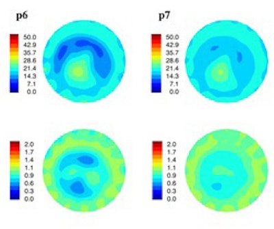
 Images acquired by microwave tomography system demonstrate electrical property distribution of a cancer patient's left breast, taken somewhat into her therapy. The breast outline and the features inside the breast that relate to fibroglandular tissue inside the adipose tissue can be visualized. This breast has undergone treatment and so most of the tumor has disappeared. Image courtesy of Neil Epstein and the University of Calgary.
While the microwave technique does not have the spatial resolution of mammography, it could offer better specificity, giving clinicians the ability to determine whether a lesion is malignant or benign, according to the researchers.