Dear AuntMinnie Member,
How to manage lesions characterized as BI-RADS 3 is one of the trickiest dilemmas breast imaging specialists face. But new research suggests that these probably benign lesions may not need to be monitored as frequently as previously thought.
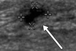
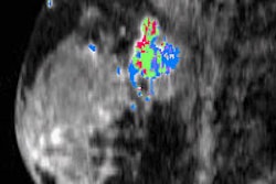
Researchers analyzed data from the American College of Radiology Imaging Network (ACRIN) 6666 trial to assess how often lesions detected on ultrasound and categorized as BI-RADS 3 turn out to be malignant. Current guidelines call for these lesions to be monitored for suspicious changes starting at six months.
But of the total 745 BI-RADS 3 lesions, only one turned out to have suspicious changes at six-month follow-up; at 12 months, one additional lesion turned out to be malignant. Learn more about the implications of the study by clicking here, or visit our Women's Imaging Digital Community at women.auntminnie.com.
Women and minorities in radiology
Should radiology be doing more to recruit women and minorities? That's the suggestion of a new study in our Residents Digital Community that indicates the percentage of women and minorities in radiology has barely budged over the past eight years.
The researchers found that while the number of women and minority medical students is approaching levels found in the population, they don't appear to be picking radiology as a specialty. The finding echoes recent studies indicating that women appear to be shunning radiology.
What do the findings portend for radiology? And can the specialty do a better job of recruiting women and minorities? Read more by clicking here, or visit the community at residents.auntminnie.com.
Finally, does training emergency physicians on the appropriate time to order CT pulmonary angiography (CTPA) exams to detect pulmonary embolism reduce inappropriate utilization? Not necessarily, according to Texas researchers.
The group found that emergency docs who got the training actually ended up ordering more CTPA studies. Find out why the boomerang effect may have occurred by clicking here.