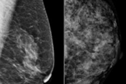
Women's imaging vendor Hologic said it has received approval from the U.S. Food and Drug Administration (FDA) for its C-View 2D imaging software, to be used with its Selenia Dimensions 3D breast tomosynthesis system.
C-View 2D images may now be used in place of the conventional 2D exposure previously required as part of a Hologic breast tomosynthesis screening exam, according to the firm.
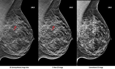
The software generates 2D images from the 3D data acquired during a breast tomosynthesis exam, eliminating the need for additional 2D exposures and, therefore, reducing radiation dose and exam time, Hologic said.
Left, 3D tomosynthesis image slice; middle, C-View 2D image; right, conventional 2D image. Images courtesy of Hologic."Large-scale clinical studies have shown that screening with Hologic's 3D mammography technology allows radiologists to visualize the breast in greater detail than with 2D mammography alone, which results in earlier detection of cancers while at the same time reducing the false positives associated with conventional 2D mammography that cause unnecessary anxiety and cost," the company said in a statement.



![A normal mammogram confirmed by three-year radiologic follow-up illustrates reader-marked regions of interest (ROIs) during (A) unaided (round 1) and (B) artificial intelligence (AI)–assisted (round 2) reading. Each colored dot represents an ROI for recall by a human reader. Readers could mark more than one ROI per case, represented by multiple dots of the same color. During AI-assisted reading, the AI system displayed three visible prompts: two with suspicion of malignancy scores of 35% (left mediolateral oblique [L MLO] and craniocaudal [L CC]) and one with a suspicion of malignancy score of 10% (right craniocaudal [R CC]), shown as polygonal overlays. Without AI, six of 10 readers (60%) marked a false-positive ROI. With AI assistance, this fell to two of 10 (20%). R MLO = right mediolateral oblique.](https://img.auntminnie.com/mindful/smg/workspaces/default/uploads/2026/07/2026-07-14-radiology-mammogram-ai-auto-bias.H0bYO8QlWs.jpg?auto=format%2Ccompress&fit=crop&h=100&q=70&w=100)


![A normal mammogram confirmed by three-year radiologic follow-up illustrates reader-marked regions of interest (ROIs) during (A) unaided (round 1) and (B) artificial intelligence (AI)–assisted (round 2) reading. Each colored dot represents an ROI for recall by a human reader. Readers could mark more than one ROI per case, represented by multiple dots of the same color. During AI-assisted reading, the AI system displayed three visible prompts: two with suspicion of malignancy scores of 35% (left mediolateral oblique [L MLO] and craniocaudal [L CC]) and one with a suspicion of malignancy score of 10% (right craniocaudal [R CC]), shown as polygonal overlays. Without AI, six of 10 readers (60%) marked a false-positive ROI. With AI assistance, this fell to two of 10 (20%). R MLO = right mediolateral oblique.](https://img.auntminnie.com/mindful/smg/workspaces/default/uploads/2026/07/2026-07-14-radiology-mammogram-ai-auto-bias.H0bYO8QlWs.jpg?auto=format%2Ccompress&fit=crop&h=112&q=70&w=112)