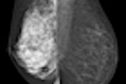
The American Society of Breast Disease (ASBD) has issued a statement to help physicians notify women of their breast density status and how it affects their lifetime cancer risk.
"If women know their lifetime risk ... and whether or not their breasts are dense, they will be better equipped to understand the recommendations their physicians make -- and make more informed screening decisions," the society said.
ASBD noted the following in its statement:
- Effective screening for breast cancer is not just a single test, but a multidisciplinary, inclusive process involving primary physicians, ob/gyns, and patients.
- Because breast screening is multidisciplinary, the various clinicians involved may have different opinions about it.
- Technical and scientific information related to dense breast tissue should be analyzed by medical professionals.
- Information about a woman's breast tissue density should be communicated to her in a way that she can understand.
- Medical professionals should make recommendations and establish quality performance standards based on science, experience, and judgment.
- Shared decision-making between patients and their physicians leads to better patient satisfaction, compliance, and treatment outcomes.
Five states -- California, New York, Virginia, Connecticut, and Texas -- now mandate that breast density results be communicated to patients by letter, and these states include 30% of the nation's women, ASBD said. Similar legislation is being discussed in more than a dozen other states, and Congress is considering a federal law.