Dear Women's Imaging Insider,
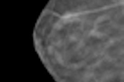
The issue of breast tissue density and whether it's a cancer risk factor has been in the news lately, especially due to grassroots advocacy efforts across the U.S. to establish breast density notification laws.
Conventional wisdom dictates that it's primarily younger women who have dense breast tissue. But breast density can vary regardless of a woman's age. Because of this, screening decisions shouldn't necessarily be linked to breast tissue density, according to researchers at Albert Einstein Medical Center in Philadelphia. Click here to read what they suggest instead.
We're also highlighting coverage of a May 23 webinar presented by Frost & Sullivan that discussed how breast density notification legislation will influence both clinical practice and the adoption of new technology in the next five years. Click here to read more.
Once you've read these stories, take a look at what else is going on in the Women's Imaging Digital Community:
- Read why breast MRI helps predict chemotherapy's effectiveness and how BI-RADS categories for breast MRI work well in predicting malignancies.
- Discover why researchers believe that accelerated partial-breast irradiation brachytherapy is an effective treatment for patients diagnosed with ductal carcinoma in situ.
- Check out our coverage of the American Society of Breast Surgeons annual meeting to find out whether radiotherapy after mastectomy is really beneficial and whether infrared thermography is ready for prime time.
- Peruse some of our coverage of the American Roentgen Ray Society meeting to discover why "no family history" is no reason to deny breast cancer screening, and you can also learn just how much digital tomography cuts recall rates.
As always, if you have a comment, report, or article idea to share about any aspect of women's imaging, I invite you to contact me.