Wednesday, December 2 | 3:40 p.m.-3:50 p.m. | SSM01-05 | Arie Crown Theater
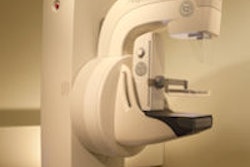
Digital mammography shortens acquisition and interpretation time for diagnostic exams, according to this scientific session presentation to be given by researchers from the University of North Carolina School of Medicine in Chapel Hill.Dr. Cherie Kuzmiak and colleagues performed the study to track their own center's transition from film-screen to soft-copy digital mammography. They included 295 patients in the project.
"We're in the process of converting to digital mammography with soft-copy display in our diagnostic breast center, and we wanted to see how this would affect our daily workflow and patient throughput," Kuzmiak told AuntMinnie.com.
Kuzmiak's team conducted the study in three phases: first, measuring technologist acquisition and processing times, and radiologist interpretation time for patients imaged with film-screen mammography; second, measuring these same times for patients imaged with a digital radiography (DR)-based mammography system, with interpretations done on a soft-copy display; and third, the same measurements three months after the soft-copy display had been installed for patients again imaged with the DR unit.
Diagnostic mammography acquisition time with processing was 2.68 minutes per image for film-screen in phase 1, 1.55 minutes per image for digital in phase 2, and 1.82 minutes per image for digital in phase 3. The mean interpretation time was 1.17 minutes per image for film-screen mammography in phase 1, 42 seconds per image for digital in phase 2, and 43 seconds per image for digital in phase 3.
Kuzmiak's team was surprised to find that interpretation times for diagnostic mammograms decreased with digital mammography and soft-copy display as compared to film-screen, Kuzmiak said.
"Published literature on screening mammography has shown increased interpretation times, so we were surprised to find that the interpretation times for diagnostic, digital mammograms as compared to film mammograms decreased," she said. "We did expect the decrease in acquisition time, since with DR full-field digital mammography processing time is completely eliminated."