Monday, November 30 | 3:30 p.m.-3:40 p.m. | SSE02-04 | Room E450A
Conebeam breast CT, when used with and without contrast, shows promise in the diagnostic workup of breast lesions, according to researchers from Elizabeth Wende Breast Care center in Rochester, NY.Dr. Posy Seifert and colleagues compared conebeam breast CT (CBBCT)'s performance in evaluating breast lesions to full-field digital mammography (FFDM) and ultrasound. The team enrolled 33 women, all of whom had a diagnostic evaluation with physical exam and ultrasound prior to undergoing breast biopsy. A total of 26 women had CBBCT and seven women had CBBCT with contrast. Pathology reports showed 20 malignancies, 12 benign lesions, and one atypical lesion.
Seifert found that agreement between CBBCT both with and without contrast and FFDM exams was 94%; the agreement between the technology and ultrasound was 76%. CBBCT found 97% of the lesions that had been confirmed via pathology.
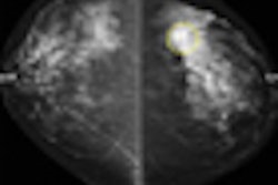
CBBCT used with contrast found new lesions that hadn't been previously seen with FFDM or ultrasound in 29% of the study cohort. CBBCT without contrast visualized lesions with better conspicuity than the other two modalities: In 82% of the included cases, CBBCT, both with and without contrast, provided more information on the extent of disease.
Seifert conceded that although CBBCT shows promise as a tool for the diagnostic workup of breast lesions, further studies are necessary, as this one had a small patient cohort.