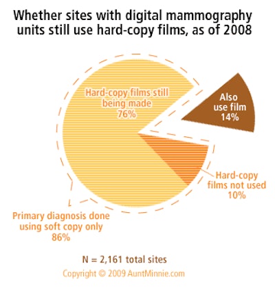
Of the sites that have digital mammography units, 86% indicate that primary diagnosis is done using soft copy only (e.g., filmless, done using the image monitor) and 14% say that primary diagnosis is also done using film. This 86% is comprised of 76% that indicate hard-copy films are made for other purposes at least some of the time, and 10% that indicate hard-copy films are not used at all. The top two purposes for which hard copy films are made are to "provide films to referral physicians" and to "provide films to patients."

Based on responses to IMV's 2008 Mammography Center Survey of U.S. Hospitals and Nonhospitals.
AuntMinnie's IMV MarketStat is provided to AuntMinnie.com by IMV Medical Information Division, Inc. of Des Plaines, IL.

Click here to buy complete IMV Market Reports
MarketStat ArchivesDistribution of mobile vs. fixed PET imaging sites
Nuclear medicine -- Average waiting time
Radiation oncology -- Types of images used in radiation therapy treatment plans
Vascular MRI -- Percent of sites performing and percent of MRI procedures
Single versus multislice detectors in CT installed base
Budgets for nuclear medicine radiopharmaceuticals
Formats used for images sent or received by radiation oncology departments
MRI adult versus pediatric patient visit mix
Cath lab device budgets for 2006
Copyright © 2009 AuntMinnie.com

![A normal mammogram confirmed by three-year radiologic follow-up illustrates reader-marked regions of interest (ROIs) during (A) unaided (round 1) and (B) artificial intelligence (AI)–assisted (round 2) reading. Each colored dot represents an ROI for recall by a human reader. Readers could mark more than one ROI per case, represented by multiple dots of the same color. During AI-assisted reading, the AI system displayed three visible prompts: two with suspicion of malignancy scores of 35% (left mediolateral oblique [L MLO] and craniocaudal [L CC]) and one with a suspicion of malignancy score of 10% (right craniocaudal [R CC]), shown as polygonal overlays. Without AI, six of 10 readers (60%) marked a false-positive ROI. With AI assistance, this fell to two of 10 (20%). R MLO = right mediolateral oblique.](https://img.auntminnie.com/mindful/smg/workspaces/default/uploads/2026/07/2026-07-14-radiology-mammogram-ai-auto-bias.H0bYO8QlWs.jpg?auto=format%2Ccompress&fit=crop&h=100&q=70&w=100)


![A normal mammogram confirmed by three-year radiologic follow-up illustrates reader-marked regions of interest (ROIs) during (A) unaided (round 1) and (B) artificial intelligence (AI)–assisted (round 2) reading. Each colored dot represents an ROI for recall by a human reader. Readers could mark more than one ROI per case, represented by multiple dots of the same color. During AI-assisted reading, the AI system displayed three visible prompts: two with suspicion of malignancy scores of 35% (left mediolateral oblique [L MLO] and craniocaudal [L CC]) and one with a suspicion of malignancy score of 10% (right craniocaudal [R CC]), shown as polygonal overlays. Without AI, six of 10 readers (60%) marked a false-positive ROI. With AI assistance, this fell to two of 10 (20%). R MLO = right mediolateral oblique.](https://img.auntminnie.com/mindful/smg/workspaces/default/uploads/2026/07/2026-07-14-radiology-mammogram-ai-auto-bias.H0bYO8QlWs.jpg?auto=format%2Ccompress&fit=crop&h=112&q=70&w=112)