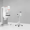
One of the limits of mammography is that it takes 2D images of a 3D structure -- hence the need to compress the tissue as much as possible so that less is hidden from the x-ray. Women with dense breast tissue face a higher risk of occult cancers being missed.
As digital mammography has come into mainstream clinical practice, stereoscopic digital mammography has become a practical possibility, according to Dr. Carl D'Orsi, director of Emory University Hospital's Breast Imaging Center in Atlanta, and colleague Dr. Mary Newell, assistant director of breast imaging. D'Orsi and Newell have been researching the efficacy of stereo mammograms along with partner BBN Technologies of Cambridge, MA, which has developed a stereo mammography workstation that allows radiologists to see 2D exams in 3D.
"There's a downside to mammography for women with dense breasts," Newell said. "Superimposed tissue can lead to false findings, or hide cancer. This stereo mammogram technology is a cost-effective way to make a 3D image simply using binocular human vision."
Not just smoke and mirrors
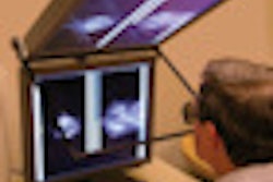
The workstation used in this research consists of a prototype stereo display, the StereoMirror SD2250 (developed by Planar Systems of Beaverton, OR), and software developed by David Getty, Ph.D., lead scientist at BBN Technologies of Cambridge, MA. The stereo display consists of two five-megapixel, grayscale display monitors mounted one above the other with an angular separation of 100° between the two.
A stereo pair of x-ray mammograms of the breast is acquired with the x-ray tube rotated by 10° between the two acquisitions while the breast remains compressed and unmoved. Each of the stereo pair images is displayed on one of two monitors, which are cross-polarized. A glass plate with a half-silvered coating is positioned between the two monitor faces, bisecting the angle between them. The image on the lower monitor is transmitted through the glass plate, while the image presented on the upper monitor is reflected from the top surface of the glass plate.
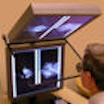
The radiologist wears passive cross-polarized glasses, allowing the left eye to see only the reflected image from the upper monitor and the right eye to see only the transmitted image from the lower monitor. The radiologist's brain fuses the two images into a single in-depth image of the internal structure of the breast. Each view of the breast can be seen in a single stereo view at full resolution or as both stereo views of both breasts simultaneously at half-resolution.
 |
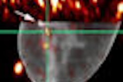
Breast images in stereo
Another technology under development for producing 3D mammography images is breast tomosynthesis, which produces a sequence of 2D slices that represents the entire volume. BBN's stereo workstation produces the same result, and its only expense is the workstation, according to Getty, who has been working on the development of the system for 14 years. He started the project by doing research with breast biopsy specimens, taking multiple stereo views of them before they went for pathology review.
"Human vision has evolved to see depth using stereoscopic vision," Getty said. "We found there was a lot of information in the stereo view that it was impossible to imagine from 2D images."
Getty presented results from the Emory study at the 2008 International Workshop on Digital Mammography in Tucson, AZ. For the trial, 1,487 women were enrolled, and each woman received a standard digital screening mammogram and a stereoscopic screening exam in a single visit. The standard exam was done with a Senographe 2000D system (GE Healthcare, Chalfont St. Giles, U.K.), and the stereoscopic exam was performed on a research Senographe 2000D with modified x-ray collimation. Five radiologist readers participated in the trial, and standard and stereo digital mammograms for each patient were read independently by two different radiologists.
The study found a reduction in false positives in the stereo mammograms of 46% compared to standard mammography, as well as an improvement of 23% in true lesion detection, most of which were calcification clusters, Getty said. False negatives were reduced by 37%.
The device is not yet cleared for use by the U.S. Food and Drug Administration, Getty said. Clearance could be fairly simple because both of the monitors used with the device have been approved for soft-copy mammography reading. In any case, the technology could have wide-ranging use once it hits the market, according to Getty.
"I see this largely as a screening tool that could dramatically cut down false positives," he said. "But it could have a place in diagnosis as well, not just for women with dense breast tissue, but for all women."
By Kate Madden Yee
AuntMinnie.com staff writer
August 29, 2008
Related Reading
SPECT/CT mammography system produces first patient images, July 30, 2008
Duke MMIL designs novel SPECT/CT system for 3D breast imaging, July 18, 2007
British firm Dexela focuses on 3D mammography workstation, October 27, 2006
Expanded role looms for 3D in breast imaging, April 11, 2006
Copyright © 2008 AuntMinnie.com