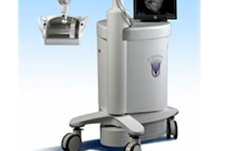
Computer-aided detection (CAD) developer R2 Technology has introduced version 8.1 of its ImageChecker mammography CAD system.
The new release offers separate operating points -- between the system's highest sensitivity and lowest false-marker rates -- for masses and microcalcifications, according to the Sunnyvale, CA-based firm. Other features include R2 Malc mark, which indicates a mass with microcalifications; EmphaSize variable size CAD marks, which correlate to lesion significance; and GreenLight user interface, which enables technologists to process mammography cases quickly and monitor film scanning from a distance, according to R2.
By AuntMinnie.com staff writers
November 21, 2005
Related Reading
Road to RSNA, R2 Technology, November 17, 2005
R2 signs three-year deal with Consorta, November 8, 2005
Road to RSNA, R2 Technology, November 7, 2005
Sectra, R2 expand partnership, August 24, 2005
R2 adds pulmonary artery patency exam tool, June 16, 2005
Copyright © 2005 AuntMinnie.com

![A normal mammogram confirmed by three-year radiologic follow-up illustrates reader-marked regions of interest (ROIs) during (A) unaided (round 1) and (B) artificial intelligence (AI)–assisted (round 2) reading. Each colored dot represents an ROI for recall by a human reader. Readers could mark more than one ROI per case, represented by multiple dots of the same color. During AI-assisted reading, the AI system displayed three visible prompts: two with suspicion of malignancy scores of 35% (left mediolateral oblique [L MLO] and craniocaudal [L CC]) and one with a suspicion of malignancy score of 10% (right craniocaudal [R CC]), shown as polygonal overlays. Without AI, six of 10 readers (60%) marked a false-positive ROI. With AI assistance, this fell to two of 10 (20%). R MLO = right mediolateral oblique.](https://img.auntminnie.com/mindful/smg/workspaces/default/uploads/2026/07/2026-07-14-radiology-mammogram-ai-auto-bias.H0bYO8QlWs.jpg?auto=format%2Ccompress&fit=crop&h=100&q=70&w=100)


![A normal mammogram confirmed by three-year radiologic follow-up illustrates reader-marked regions of interest (ROIs) during (A) unaided (round 1) and (B) artificial intelligence (AI)–assisted (round 2) reading. Each colored dot represents an ROI for recall by a human reader. Readers could mark more than one ROI per case, represented by multiple dots of the same color. During AI-assisted reading, the AI system displayed three visible prompts: two with suspicion of malignancy scores of 35% (left mediolateral oblique [L MLO] and craniocaudal [L CC]) and one with a suspicion of malignancy score of 10% (right craniocaudal [R CC]), shown as polygonal overlays. Without AI, six of 10 readers (60%) marked a false-positive ROI. With AI assistance, this fell to two of 10 (20%). R MLO = right mediolateral oblique.](https://img.auntminnie.com/mindful/smg/workspaces/default/uploads/2026/07/2026-07-14-radiology-mammogram-ai-auto-bias.H0bYO8QlWs.jpg?auto=format%2Ccompress&fit=crop&h=112&q=70&w=112)