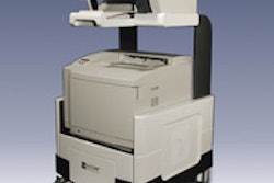
CT laser mammography (CTLM) developer Imaging Diagnostic Systems (IDSI) in a letter to shareholders today said that it is altering its course for obtaining U.S. Food and Drug Administration premarket approval (PMA) for its CTLM device.
The Plantation, FL-based vendor said that its clinical cases for PMA submission dated back to 2001, and were performed on earlier iterations of the technology. As such, it believes that the studies do not best represent the capabilities of the current device. It said it now intends to collect data using its latest systems because it believes the results will yield a stronger study to support the PMA application.
In other news, IDSI reported that it has sold a CTLM system to its Rome-based distributor Biomedical International for installation at the Hospital Civile di Teramo in Teramo, Italy.
By AuntMinnie.com staff writers
January 27, 2005
Related Reading
Imaging Diagnostic books international orders, January 20, 2005
Imaging Diagnostic nets Turkish distributor, January 4, 2005
Imaging Diagnostic gets CTLM clearance in Canada, November 14, 2003
IDSI PMA judged suitable for filing, June 18, 2003
Imaging Diagnostic Systems receives 12th U.S. patent, May 29, 2003
Copyright © 2005 AuntMinnie.com

![A normal mammogram confirmed by three-year radiologic follow-up illustrates reader-marked regions of interest (ROIs) during (A) unaided (round 1) and (B) artificial intelligence (AI)–assisted (round 2) reading. Each colored dot represents an ROI for recall by a human reader. Readers could mark more than one ROI per case, represented by multiple dots of the same color. During AI-assisted reading, the AI system displayed three visible prompts: two with suspicion of malignancy scores of 35% (left mediolateral oblique [L MLO] and craniocaudal [L CC]) and one with a suspicion of malignancy score of 10% (right craniocaudal [R CC]), shown as polygonal overlays. Without AI, six of 10 readers (60%) marked a false-positive ROI. With AI assistance, this fell to two of 10 (20%). R MLO = right mediolateral oblique.](https://img.auntminnie.com/mindful/smg/workspaces/default/uploads/2026/07/2026-07-14-radiology-mammogram-ai-auto-bias.H0bYO8QlWs.jpg?auto=format%2Ccompress&fit=crop&h=100&q=70&w=100)


![A normal mammogram confirmed by three-year radiologic follow-up illustrates reader-marked regions of interest (ROIs) during (A) unaided (round 1) and (B) artificial intelligence (AI)–assisted (round 2) reading. Each colored dot represents an ROI for recall by a human reader. Readers could mark more than one ROI per case, represented by multiple dots of the same color. During AI-assisted reading, the AI system displayed three visible prompts: two with suspicion of malignancy scores of 35% (left mediolateral oblique [L MLO] and craniocaudal [L CC]) and one with a suspicion of malignancy score of 10% (right craniocaudal [R CC]), shown as polygonal overlays. Without AI, six of 10 readers (60%) marked a false-positive ROI. With AI assistance, this fell to two of 10 (20%). R MLO = right mediolateral oblique.](https://img.auntminnie.com/mindful/smg/workspaces/default/uploads/2026/07/2026-07-14-radiology-mammogram-ai-auto-bias.H0bYO8QlWs.jpg?auto=format%2Ccompress&fit=crop&h=112&q=70&w=112)