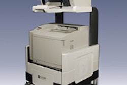
Mammography archive provider i3Archive has recently installed its breast image archiving product, National Digital Mammography Archive (NDMA), at the Philadelphia-based University of Pennsylvania Health System.
NDMA is a digital mammography storage archive that involves the use of grid computing, in which computers are distributed over multiple locations across a region, according to the Berwyn, PA-based i3Archive. The product is available through a per-case ASP solution, as well as through the Extended Local Archive Option (ELAO) system created for facilities that want NDMA services, but simultaneously maintain onsite storage, the company said.
By AuntMinnie.com staff writers
January 5, 2005
Related Reading
Road to RSNA, i3Archive, November 10, 2004
i3Archive signs up first center, May 26, 2004
GE, i3Archive partner up, January 22, 2004
iCAD, i3Archive team up, November 19, 2003
Siemens, i3Archive ink deal, September 29, 2003
Copyright © 2005 AuntMinnie.com

![A normal mammogram confirmed by three-year radiologic follow-up illustrates reader-marked regions of interest (ROIs) during (A) unaided (round 1) and (B) artificial intelligence (AI)–assisted (round 2) reading. Each colored dot represents an ROI for recall by a human reader. Readers could mark more than one ROI per case, represented by multiple dots of the same color. During AI-assisted reading, the AI system displayed three visible prompts: two with suspicion of malignancy scores of 35% (left mediolateral oblique [L MLO] and craniocaudal [L CC]) and one with a suspicion of malignancy score of 10% (right craniocaudal [R CC]), shown as polygonal overlays. Without AI, six of 10 readers (60%) marked a false-positive ROI. With AI assistance, this fell to two of 10 (20%). R MLO = right mediolateral oblique.](https://img.auntminnie.com/mindful/smg/workspaces/default/uploads/2026/07/2026-07-14-radiology-mammogram-ai-auto-bias.H0bYO8QlWs.jpg?auto=format%2Ccompress&fit=crop&h=100&q=70&w=100)


![A normal mammogram confirmed by three-year radiologic follow-up illustrates reader-marked regions of interest (ROIs) during (A) unaided (round 1) and (B) artificial intelligence (AI)–assisted (round 2) reading. Each colored dot represents an ROI for recall by a human reader. Readers could mark more than one ROI per case, represented by multiple dots of the same color. During AI-assisted reading, the AI system displayed three visible prompts: two with suspicion of malignancy scores of 35% (left mediolateral oblique [L MLO] and craniocaudal [L CC]) and one with a suspicion of malignancy score of 10% (right craniocaudal [R CC]), shown as polygonal overlays. Without AI, six of 10 readers (60%) marked a false-positive ROI. With AI assistance, this fell to two of 10 (20%). R MLO = right mediolateral oblique.](https://img.auntminnie.com/mindful/smg/workspaces/default/uploads/2026/07/2026-07-14-radiology-mammogram-ai-auto-bias.H0bYO8QlWs.jpg?auto=format%2Ccompress&fit=crop&h=112&q=70&w=112)