Optical breast imaging developer DOBI Medical Systems has named Lim Cheung as vice president of engineering. Cheung comes to the Mahwah, NJ-based firm from Trex Medical, where he served as vice president of research and development and led the development effort for that firm's digital mammography system, according to DOBI. At DOBI, Cheung will head the development team responsible for designing and testing the company's work-in-progress dynamic optical breast imaging (DOBI) system.
By AuntMinnie.com staff writersMay 18, 2000

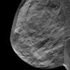

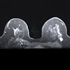





![A normal mammogram confirmed by three-year radiologic follow-up illustrates reader-marked regions of interest (ROIs) during (A) unaided (round 1) and (B) artificial intelligence (AI)–assisted (round 2) reading. Each colored dot represents an ROI for recall by a human reader. Readers could mark more than one ROI per case, represented by multiple dots of the same color. During AI-assisted reading, the AI system displayed three visible prompts: two with suspicion of malignancy scores of 35% (left mediolateral oblique [L MLO] and craniocaudal [L CC]) and one with a suspicion of malignancy score of 10% (right craniocaudal [R CC]), shown as polygonal overlays. Without AI, six of 10 readers (60%) marked a false-positive ROI. With AI assistance, this fell to two of 10 (20%). R MLO = right mediolateral oblique.](https://img.auntminnie.com/mindful/smg/workspaces/default/uploads/2026/07/2026-07-14-radiology-mammogram-ai-auto-bias.H0bYO8QlWs.jpg?auto=format%2Ccompress&dpr=2&fit=crop&h=167&q=70&w=250)


