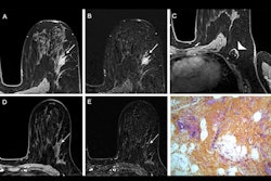
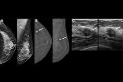
Breast MRI is effective for measuring breast tumors before a woman undergoes surgery, according to a study published June 16 in the Annals of Surgical Oncology.
"[We found that] MRI demonstrated the highest concordance with tumor size and T stage," wrote a team led by Abigail Daly, MD, of Massachusetts General Hospital in Boston.
Accurate preoperative imaging of breast tumor size is essential, as small measurement differences can influence the treatment strategy, the researchers noted. They investigated the accuracy of tumor size estimation by mammography, ultrasound, and MRI compared with pathology results.
Their study included 460 patients with breast cancer who were treated between 2019 and 2024. The team considered imaging measurements concordant if they fell within ± 20% of the pathological size of the tumor. Average tumor size on pathology was 17.3 mm.
Overall, the group found that breast MRI had the highest concordance with pathology results for measuring tumor size.
Tumor size estimation by imaging modality | |
Modality | Concordance with pathology results |
| MRI | 62% |
| Mammography | 57% |
| Ultrasound | 53% |
The researchers also found that MRI slightly overestimated tumor size (18.4 mm), while mammography and ultrasound underestimated (14.8 mm and 14.3 mm, respectively). They reported the following:
- Of all three imaging modalities, MRI showed the highest accuracy in T-stage classification (89%).
- MRI's concordance with pathology was highest for masses without nonmass enhancement.
- The modality's accuracy improved in tumors greater than 15 mm (odds ratio [OR], 2.47, with 1 as reference) and high-grade tumors (OR, 1.75), but declined in extremely dense breasts (OR, 0.42).
Despite MRI's performance, using other modalities with it to assess tumor size before surgery could be a good idea, according to the authors.
"A combined imaging approach using MRI and ultrasound may enhance preoperative size estimation," they concluded.
The complete study can be found here.