Dear AuntMinnie Member,
The Ides of March finds radiology smack in the middle of conference season, and with this issue of the Letter from the Editor we bring you reporting from the Breast Course 2007, a multidisciplinary meeting on breast health.
Staff writer Shalmali Pal is onsite in Key Largo, FL, to report on the meeting for our Women's Imaging Digital Community. Topics at the conference range from breast MRI to ductal sonography to imaging's role in the coming era of molecular testing.
In one presentation, breast MRI specialist Dr. Steven Harms reports on a new breast MRI technique that he believes has superior performance in detecting ductal carcinoma in situ (DCIS). The technique provides the greater resolution and contrast needed for DCIS detection, he believes. Get more details by clicking here.
DCIS was also the subject of a talk that touched on the impact that genetic testing and molecular markers will play in evaluation of the pathology. While some panel participants see a role, others believe that mammography will always remain the gold standard due to its value in morphology assessment. See what they said by clicking here.
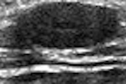
Finally, Dr. Thomas Stavros discusses the use of ultrasound as a tool to potentially redirect patients from more invasive galactography exams. But the technique needs to be in the right hands in order to be used most effectively. Learn about how he does it by clicking here.
Get these stories and more news about the world of breast imaging in our Women's Imaging Digital Community, at women.auntminnie.com.