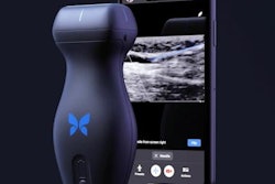
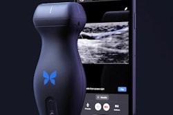
Midjourney Medical has unveiled plans for its Midjourney Scanner, a full-body ultrasound imaging device powered by 40 Butterfly Network ultrasound-on-chip imaging modules.
The scanner uses a water-immersion design in which a patient descends on a platform through a ring of ultrasonic sensors, with a target scan time of 60 seconds. The sensors send ultrasonic waves through the body from multiple angles, with the resulting data processed to produce 3D images of internal anatomy down to a fraction of a millimeter, the company said. Midjourney said the image quality is comparable to MRI but at significantly faster acquisition speeds.
Example image from the Midjourney Scanner. Each slice continuously crossfades between the raw reconstruction and its AI segmentation.Midjourney
Butterfly's imaging modules incorporate approximately 500,000 sensors and more than two petaflops of processing power.
The device is not yet cleared by the U.S. Food and Drug Administration (FDA), but Midjourney said it plans to submit regular test results to the FDA as it develops diagnostic capabilities. The company also said it currently intends to offer body composition mapping as an initial use case. It plans to open its first scanner location, integrated into a spa facility in San Francisco, in 2027.
The collaboration with Butterfly Network is enabled through Butterfly's Embedded co-development program, and Butterfly previously disclosed up to $74 million in expected payments over a five-year term in a November 2025 filing with the U.S. Securities and Exchange Commission (SEC). Midjourney said its longer-term goal is to deploy more than 50,000 scanners worldwide by 2031.








![Examples of ultrasound findings and techniques. (A) Images in a 39-year-old male patient with a mass in the left thigh. The mass is heterogeneous on the B-mode US image (compared with the patient in D) and showed increased microvascularity (superb microvascular imaging [SMI]) and shear-wave elastography (SWE) values. Undifferentiated pleomorphic sarcoma was diagnosed at biopsy (with pleomorphic rhabdomyosarcoma in surgical specimen). (B) Images in an 18-year-old male patient with a mass in the left leg. The mass is hypoechoic on the B-mode image, with no other findings suggestive of malignancy. The lesion is in contact with the cortex of the tibia, which is slightly irregular. CT revealed a doubtful anteromedial tibial erosion. The microvascular study demonstrated high vascularization, suggestive of malignancy. Periosteal Ewing sarcoma was diagnosed with both histologic and immunohistochemical confirmation. (C) Images in a 69-year-old female patient with a lump growing on the outside of the left leg. Multiple SWE examinations were performed (please note the high values obtained in the measurements, whereas the color map highlights the stiffness relative to adjacent tissues). SMI showed areas of increased vascularization to target for sampling. Undifferentiated spindle cell sarcoma was diagnosed at biopsy, with residual leiomyosarcoma in the surgical specimen after neoadjuvant therapy. (D) Images in a 56-year-old female patient with a mass in the right thigh. The mass is heterogeneous at both B-mode ultrasound (similar to patient A) and MRI (coronal T2-weighted spectral attenuated inversion recovery [SPAIR]; T1-weighted pre-contrast and postcontrast imaging), which even shows uptake after the administration of paramagnetic contrast material, which is traditionally suggestive of malignancy. Low values at SMI and elastography are suggestive of benignity. Spindle cell lipoma was diagnosed at biopsy, with atypical spindle cell lipomatous tumor in the surgical specimen.](https://img.auntminnie.com/mindful/smg/workspaces/default/uploads/2026/08/images-radiol250278fig2.APCFLSvX6p.jpg?auto=format%2Ccompress&fit=crop&h=112&q=70&w=112)


