
The presence of an echogenic rind surrounding suspicious breast masses on ultrasound may serve as a more specific sign of malignancy, according to research published September 17 in the American Journal of Roentgenology.
Echogenic rinds are thick hyperechoic bands that surround a mass, often disrupting the texture of surrounding tissue. They can be found most often in invasive ductal and lobular carcinomas, wrote a team led by Derek Nguyen, MD, from Duke University in Durham, NC. The group noted that including the rind in size measurements reduces tumor underestimation compared to surgical pathology.
“These results provide insights into the clinical implications of an echogenic rind on ultrasound,” the Nguyen team wrote.
The BI-RADS 6th edition is expected to include language related to breast ultrasound, which the team highlighted will include an echogenic rind as a new associated feature.
Nguyen and colleagues evaluated whether including this feature improves breast malignancy diagnosis. They also studied associations between the pathologic tumor characteristics of an echogenic rind and a suspicious mass on breast ultrasound.
Using a retrospective data set, the researchers included 511 women with an average age of 52 in their study. The women had 583 suspicious masses in total (BI-RADS 4 or 5), 249 of which were diagnosed as breast malignancy.
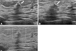
 Diagnostic transverse ultrasound image of the left breast in a 64-year-old woman shows irregular hypoechoic nonparallel mass with indistinct margins corresponding to palpable finding, assessed as BI-RADS category 4. Both readers assessed mass as having associated echogenic rind with complete circumferential extent (arrowheads). Ultrasound-guided biopsy of mass yielded diagnosis of invasive ductal carcinoma (intermediate grade, luminal A molecular subtype, no lymphovascular invasion).ARRS
Two radiologists independently reviewed ultrasound examinations for echogenic rinds (requiring ≥25% circumferential extent) associated with masses. They also measured masses with and without the inclusion of echogenic rinds.
The presence of an echogenic rind yielded high specificity values, including 81% and 85% for readers one and two, respectively. However, it also led to modest sensitivity, 58% for reader one and 51% for reader two.
The presence of an echogenic rind also showed significant associations with invasive cancer and invasive cancer molecular subtype for both readers. This feature is more often found in invasive ductal and lobular breast cancers and in HER2-enriched and luminal A subtypes.
Associations between radiologist-assessed echogenic rind, invasive cancer, and molecular subtype | ||
Cancer subtype | Reader one | Reader two |
Invasive ductal cancer | 62% | 53% |
Invasive lobular cancer | 57% | 53% |
DCIS | 24% | 18% |
Luminal A | 66% | 62% |
Luminal B | 48% | 32% |
HER2-enriched | 100% | 67% |
Basal-like | 49% | 31% |
| *All results achieved statistical significance. | ||
In 168 resected malignancies, the team assessed the difference between pathologic tumor size (average, 19 mm) and ultrasound-based mass size, without and with inclusion of echogenic rinds. For reader one, the differences were 4 mm and 3 mm, while reader two indicated differences of 7 mm and 5 mm, respectively.
The study authors highlighted that despite the results, accuracy for a diagnosis of breast malignancy “was not improved by requiring a greater circumferential extent to define the presence of an echogenic rind.”
“Interreader agreement was moderate for the presence of an echogenic rind and minimal for classification of an echogenic rind’s circumferential extent,” they added.
Still, the team recommended that radiologists should consider including any identified echogenic rind in their routine ultrasound measurements for suspicious masses.
“By more reliably delineating tumor margins and disease extent, such measurements could aid surgical planning, particularly for breast-conserving surgeries,” the group wrote.
Read the full study here.








![Examples of ultrasound findings and techniques. (A) Images in a 39-year-old male patient with a mass in the left thigh. The mass is heterogeneous on the B-mode US image (compared with the patient in D) and showed increased microvascularity (superb microvascular imaging [SMI]) and shear-wave elastography (SWE) values. Undifferentiated pleomorphic sarcoma was diagnosed at biopsy (with pleomorphic rhabdomyosarcoma in surgical specimen). (B) Images in an 18-year-old male patient with a mass in the left leg. The mass is hypoechoic on the B-mode image, with no other findings suggestive of malignancy. The lesion is in contact with the cortex of the tibia, which is slightly irregular. CT revealed a doubtful anteromedial tibial erosion. The microvascular study demonstrated high vascularization, suggestive of malignancy. Periosteal Ewing sarcoma was diagnosed with both histologic and immunohistochemical confirmation. (C) Images in a 69-year-old female patient with a lump growing on the outside of the left leg. Multiple SWE examinations were performed (please note the high values obtained in the measurements, whereas the color map highlights the stiffness relative to adjacent tissues). SMI showed areas of increased vascularization to target for sampling. Undifferentiated spindle cell sarcoma was diagnosed at biopsy, with residual leiomyosarcoma in the surgical specimen after neoadjuvant therapy. (D) Images in a 56-year-old female patient with a mass in the right thigh. The mass is heterogeneous at both B-mode ultrasound (similar to patient A) and MRI (coronal T2-weighted spectral attenuated inversion recovery [SPAIR]; T1-weighted pre-contrast and postcontrast imaging), which even shows uptake after the administration of paramagnetic contrast material, which is traditionally suggestive of malignancy. Low values at SMI and elastography are suggestive of benignity. Spindle cell lipoma was diagnosed at biopsy, with atypical spindle cell lipomatous tumor in the surgical specimen.](https://img.auntminnie.com/mindful/smg/workspaces/default/uploads/2026/08/images-radiol250278fig2.APCFLSvX6p.jpg?auto=format%2Ccompress&dpr=2&fit=crop&h=167&q=70&w=250)


