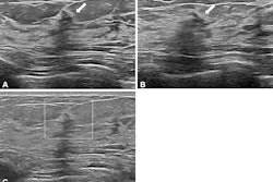
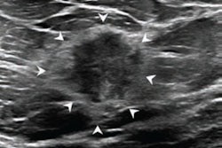
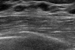
A new ultrasound approach could help distinguish fluid from solid breast masses with high accuracy, according to research published December 17 in Radiology Advances.
A team led by Muyinatu Bell, MD, from Johns Hopkins University in Baltimore, MD, reported that when generalized contrast-to-noise ratio was applied to short-lag spatial coherence images, doctors accurately identified masses 96% of the time.
“We found that our technique can distinguish complicated cysts, which are benign, from solid masses, which could be benign or malignant,” Bell told AuntMinnie.
Complicated cysts can appear on ultrasound like solid masses, which results in multiple unnecessary procedures or follow-up exams. When used on dense breasts, ultrasound waves can scatter before reaching masses, resulting in acoustic cluttering on images.
Bell and colleagues attempted to improve how ultrasound signals are processed with an approach that is “coherence-based.” This means that the image relies on how similar signals are to neighboring signals, resulting in a number score for each mass. The researchers hypothesized that this would make it easier for radiologists to find suspicious masses on ultrasound.
“Our approach is the same as the standard breast ultrasound exam,” Bell said. “The only difference is the process used to display the recorded information. We display coherence information, then apply a new metric to assess the image and determine the final result.”
The team performed a secondary analysis of the Advanced Ultrasound Signal Processing of Suspicious Breast Images (AUSPICIOUS) observational study, also being led by Johns Hopkins University. This study enrolled women from 2018 to 2023 who were scheduled for ultrasound-guided procedures or follow-up of at least one breast mass.
 Conventional ultrasound of breast tissue (top) compared to the new method (bottom). Conventional ultrasound relies on the amplitude of signals, turning high and low signals into blacks, whites, and grays. The new method is “coherence-based,” meaning the image relies on how similar signals are to neighboring signals.Johns Hopkins University
The researchers focused on a generalized contrast-to-noise ratio that was applied to short-lag spatial coherence images, with regions of interest determined by six radiologists.
Final analysis included 145 breast masses from 115 women with an average age of 52 years. This included 16 complicated cysts and 96 solid masses.
The team’s approach led to an average area under the curve (AUC) of 0.96 for complicated cyst versus solid mass characterization. Reading with B-mode images led to an average AUC of 0.67 (p < 0.05).
Interreader agreement improved from fair with B-mode (κ = 0.4) to moderate with the generalized contrast-to-noise ratio approach with a 0.76 threshold (κ = 0.59, p < 0.00001).
“The results are logical based on what I know about coherence imaging and the new [generalized contrast-to-noise ratio] metric,” Bell said. “Others might be surprised that a single signal processing change, followed by a single metric, are the only changes needed to achieve the presented performance.”
Bell also told AuntMinnie that the research team is “looking to combine our technique with light to create photoacoustic images that can further distinguish whether a solid mass is benign or malignant.”
Read the full study here.