Sound touch elastography can noninvasively evaluate liver stiffness tied to chronic heart failure, a study published January 1 in the American Journal of Cardiology found.
Researchers led by Jing-Wu Yang from Guangdong Second Provincial General Hospital in Guangzhou, China, found that patients with reduced ejection fraction and ride-sided heart failure had significantly higher liver stiffness values than control group patients.
“Sound touch elastography is a useful clinical technique … which could help patients with chronic heart failure manage their treatment regimens,” Yang and colleagues wrote.
Heart failure can affect the health of other organs. For the liver, this effect is known as cardiohepatic syndrome and includes liver congestion and hepatic artery hypoperfusion.
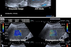
Sound touch elastography is a modified shear wave elastography method that uses multiple acoustic radiation force impulses to target an extended region of interest. The resulting display is a real-time colored stiffness map.
Yang's team studied liver damage in chronic heart failure patients using sound touch elastography. They included 150 patients in their study and divided them into distinct groups. These included patients with reduced ejection fraction (n = 45), mildly reduced ejection fraction (n = 40), right-sided heart failure (n = 25), and control (n = 40).
The group found that while patients with mildly reduced ejection fraction increased slightly compared with the control group patients, sound touch elastography showed significantly higher stiffness values in patients with reduced ejection fraction and right-sided heart failure.
| Liver stiffness values in chronic heart failure patients | ||
|---|---|---|
| Heart failure subgroups | Liver stiffness measurements | p-value (compared with control group) |
| Control | 5.4 kPa | N/A |
| Reduced ejection fraction | 8.4 kPa | < 0.05 |
| Right-sided heart failure | 10.3 kPa | < 0.05 |
| Mildly reduced ejection fraction | 5.9 kPa | > 0.05 |
Looking at other parameters, the team also reported that the right atrial volume index and diameter of the hepatic veins were significantly greater in the right-sided heart failure patients compared with the healthy controls.
Additionally, patients with reduced ejection fraction had lower left ventricular ejection fraction along with greater liver stiffness. Finally, patients with reduced ejection fraction and right-sided heart disease showed increased glutamyl transpeptidase and alkaline phosphatase values.
The study authors suggested that while sound touch elastography can detect and quantify the effect of chronic heart failure on the liver, liver fibrosis and severe liver congestion should also be considered in this setting.
“Our findings offer some reference for the early management of liver damage in patients with heart failure,” they wrote.
The full study can be found here.