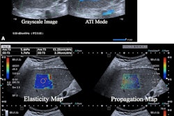
Contrast-enhanced ultrasound (CEUS) with Kupffer-cell agents has high specificity and sensitivity for diagnosing liver cancer, suggest findings published August 29 in Radiology.
Researchers led by Lingling Li, MD, from Sun Yat-Sen University Cancer Center in Guangzhou, China, found that based on CEUS LI-RADS version 2017 guidelines, sulfur hexafluoride and perfluorobutane imaged by CEUS had comparable specificity when characterizing hepatocellular carcinoma (HCC) lesions. However, ultrasound enhanced with perfluorobutane achieved significantly higher sensitivity compared with sulfur hexafluoride enhancement.
"Our results suggested that perfluorobutane-enhanced ultrasound provides an accurate noninvasive diagnosis of HCC, consistent with previous studies," Li and colleagues wrote.
Pure blood pool agents such as sulfur hexafluoride microbubbles and Kupffer-cell agents like perfluorobutane microbubbles are the two main types of contrast agents used in ultrasound. Each agent has its own distinct advantages. On one hand, sulfur hexafluoride can't cross the vascular endothelium, which means that it generates pure vascular images. On the other hand, perfluorobutane can be specifically ingested by Kupffer cells in the liver, making way for additional lesion characterization in the Kupffer phase, more than 10 minutes after contrast injection. (Lesions that don't have functioning Kupffer cells show a defect in images.)
Despite previous research showing the potential of CEUS for liver imaging, the modality has not been recommended by the American Association for the Study of Liver Diseases (AASLD). The association cites as reasons for concern potential selection and publication bias of current CEUS literature, as well as uncertainty about the generalization of study results.
Li and colleagues previously demonstrated that a modified CEUS method using perfluorobutane had high accuracy in diagnosing liver cancer. This method classifies certain liver observations as LI-RADS-5 rather than LI-RADS-4 or LI-RADS-M (probable or definite malignancy) based on Kupffer-phase findings.
Li and colleagues compared the diagnostic performance of three algorithms for HCC diagnosis in a prospective, multicenter study. The algorithms included the following: CEUS LI-RADS with sulfur hexafluoride-enhancement; CEUS LI-RADS with perfluorobutane-enhancement; and the modified algorithm that uses perfluorobutane.
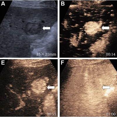
 Ultrasound shows images of a 66-year-old male at risk for HCC due to hepatitis B virus infection and liver cirrhosis. Gray-scale ultrasound was initially examined. (A) Gray-scale image shows hypoechoic observation in segment VI measuring 25 x 21 mm (arrow). The participant underwent sulfur hexafluoride-enhanced ultrasound. (B) Arterial phase image at 14 seconds after injection shows nonrim hyperenhancement (arrow). (C) Portal venous phase image at 1:04 minutes does not show early washout (arrow). (D) Late-phase image at five minutes shows mild washout (arrow). The participant underwent evaluation by perfluorobutane-enhanced ultrasound on the same day. There was a minimum interval of 30 minutes between two examinations. (E) Arterial phase image at 15 seconds after injection shows nonrim hyperenhancement (arrow). (F) Portal venous phase image at one minute does not show early washout (arrow). (G) Late-phase image at 4:59 minutes shows late and mild washout (arrow). (H) Kupffer-phase image at 20 minutes shows mild Kupffer defect. The observation was classified as LI-RADS-5 regardless of the algorithms that were used. Pathologic diagnosis based on surgical resection was HCC. Images and caption courtesy of the RSNA.
The team included data from 375 patients with an average age of 56 years with 424 ultrasound findings. Of this total, 345 were HCCs, 40 were non-HCC malignancies, and 39 were benign lesions.
The researchers found that both contrast agents based on the LI-RADS measure showed no significant difference in sensitivity and specificity. However, the modified algorithm with perfluorobutane had increased sensitivity along with a nonsignificant decrease in specificity compared to LI-RADS with sulfur hexafluoride-enhancement.
| Comparison of algorithms in HCC diagnosis* | |||
| Measure | LI-RADS perfluorobutane | LI-RADS sulfur hexafluoride | Modified algorithm with perfluorobutane |
| Specificity | 96% | 95% | 92% |
| Sensitivity | 60% | 58% | 80% |
The study authors called for future research to investigate whether these findings can be generalized to other populations. They wrote that this especially goes for results related to the modified algorithm for ultrasound enhanced with perfluorobutane.
In an accompanying editorial, Tae Kyoung Kim, MD, PhD, from the University of Toronto also highlighted the importance of validating these findings in other geographic regions. However, he also noted the potential CEUS enhanced with perfluorobutane has in improving HCC diagnosis.
"Consequently, prospective trials similar to the current study should be conducted in other geographic regions, particularly those with a higher incidence of liver cirrhosis and other risk factors," Kim wrote.
The full study can be found here.