AI assistance can enhance the performance of nonexpert clinicians on lung ultrasound exams performed in the intensive care unit (ICU), suggest findings published July 1 in Critical Care.
A team led by Phung Tran Huy Nhat from the Oxford University Clinical Research Unit in Ho Chi Minh City, Vietnam, found that accuracy, examination times, and confidence were all improved for clinicians with AI assistance.
"Developing an AI tool that can assist inexperienced users in this setting could be highly beneficial for patient outcomes and improving quality of care," Nhat and colleagues wrote.
When working in the ICU, interpreting lung ultrasound images can be challenging for clinicians. This especially goes for medical centers in low- to middle-income countries where training resources are limited.
Researchers have looked toward AI as a potential tool to help automate ultrasound imaging and assist clinicians with image interpretation. This includes its potential in emergency settings such as the ICU.
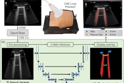
Nhat and colleagues developed a real-time AI-assisted lung ultrasound system and wanted to investigate its usefulness in a low-resource ICU. The system consists of an AI model integrated into the open-source Plugin-based Real Time UltraSound software platform (PRETUS). It provides continuous real-time prediction through a laptop and can be used in both prerecorded lung ultrasound clips and the real-time clinical environment. The system also captures the user's prediction, model prediction, time-to-interpretation, and confidence. The team also highlighted that it can be used in "any" ultrasound machine with a video output port.
For their prospective study, the authors tested and validated the AI tool in three phases. In the first phase, they evaluated the performance of four different clinical user groups in interpreting lung ultrasound clips. In the second phase, they evaluated the performance of 57 nonexpert clinicians with and without AI assistance for interpreting retrospective, offline lung ultrasound clips.
For the third phase, the team prospectively assessed the performance of 14 clinicians in the ICU. The clinicians in total examined seven patients with and without AI assistance and were interviewed afterward on using the tool.
The researchers reported that the average accuracy of novice users was 68.7% compared with 72.2% in intermediate users and 73.4% in advanced users. Ultrasound experts meanwhile had an average accuracy of 95%, significantly higher than the aforementioned users (p < 0.001).
Nhat et al found that their AI tool improved the performance of the clinicians in several areas, including accuracy, examination times, and confidence.
| Comparison of clinicians performing lung ultrasound exams with, without AI assistance | |||
| Without AI assistance | With AI assistance | p-value | |
| Average accuracy (retrospective clips) | 68.9% | 82.9% | < 0.001 |
| Average accuracy (prospective clips) | 68.1% | 93.4% | < 0.001 |
| Median time-to-interpret | 12.1 seconds | 5 seconds | < 0.001 |
| Median confidence level (out of 4) | 3 | 4 | N/A |
While clinicians who were interviewed after using the AI tool praised its usefulness and indicated interest in future use of the tool, they also raised concerns about data privacy and legal responsibility when using the tool. The study authors called their concerns "valid."
"As the application of AI in ultrasound is in its infancy, there are relatively few regulations on how to legally implement it in routine healthcare practice or who will be response for AI-derived medical errors, particularly in low resource settings," they wrote.
Still, the team suggested that based on the study's results, the AI tool could help with management of critically ill patients with lung pathologies.
"This could especially benefit the monitoring of patients during fluid resuscitation where fluid balance is critical to achieve a stable hemodynamic status without causing fluid overload," the group wrote.
The entire study can be found here.