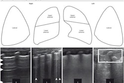
Researchers have developed lung ultrasound cutoff scores that categorize the severity of pneumonia for COVID-19, according to a study published March 10 in Medicina Clínica.
The team, led by Jaime Gil-Rodríguez from San Cecilio University Hospital in Spain, found that lung ultrasound is a good predictor of poor outcome and 28-day mortality in COVID-19 patients.
"This categorization will make it possible to establish an early prognosis for the patient, and to guide the most appropriate treatment on an individual basis," Gil-Rodríguez and colleagues wrote.
Lung ultrasound's usefulness has been explored in COVID-19 protocols, the investigators wrote, highlighting it as an "easy-to-learn, nonionizing and repeatable technique." However, they also noted that no benchmark points have been addressed as optimal cutoff points for determining the severity of the pneumonia. Gil-Rodríguez and colleagues conducted a literature review to establish the best cutoff points based on lung ultrasound findings.
They examined data from 11 studies published in 2020 and 2021 which included a total of 1,308 hospitalized patients with COVID-19 pneumonia. They used a 0- to 30-point scoring system for lung ultrasound findings, with 30 indicating high severity.
Among the cutoff points proposed in the studies, only the lung ultrasound score of greater than 15 could be validated for its original endpoint. The researchers reported that this cutoff score also showed the strongest relation with poor outcome, with an odds ratio (OR) of 3.63 (with 1 as reference).
The team also examined lung ultrasound scores with its own cohort of 127 patients, finding that lung ultrasound was statistically associated with poor outcome (OR = 1.30) and with 28-day mortality (OR = 1.02). They reported that a cutoff score of 15 showed the best diagnostic performance in differentiating between mild and severe pneumonia, with an area under the curve (AUC) value of 0.65.
Finally, while a lung ultrasound score of seven or less showed high sensitivity (89%) in ruling out poor outcome in the data from the 11 studies, a score of greater than 20 showed high specificity (86%) in predicting poor outcome. Although these same measures trended upward but did not achieve statistical significance in the researchers' own cohort of 127 patients, which the team suggested could be because of limited sample size or due to patients dying of comorbidities rather than pneumonia.
The study findings could help clinicians better predict COVID-19 outcomes, as a lung ultrasound score of seven and under can indicate mild pneumonia, while one above 20 likely indicates severe pneumonia, the team concluded.