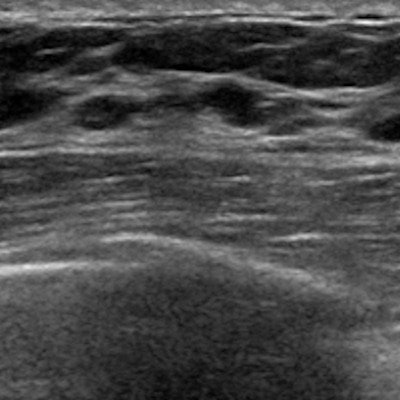
Axillary scanning in women undergoing a diagnostic breast ultrasound has minimal impact on cancer detection, according to findings published April 6 in Clinical Imaging.
A team led by Dr. Iris Chen from the University of California, Los Angeles found that less than 1% of incidental axillary findings were given a BI-RADS category 3 or 4, none of which resulted in the diagnosis of an occult breast cancer. In other words, such scanning of the axilla may not be needed.
"Omitting routine scanning of the axilla for all diagnostic breast ultrasounds may save time and reduce patient anxiety related to unnecessary follow-up and biopsies, without adversely affecting patient care," Chen and co-authors wrote.
Breast ultrasound is used in women undergoing diagnostic and screening breast imaging. However, the researchers noted that it can have high false-positive rates and can be time-consuming to perform.
While scanning the axilla is good for assessing axillary lymphadenopathy, it may be merely optional in women with a low likelihood of breast cancer. One side effect of the COVID-19 vaccine and booster is the presence of axillary lymphadenopathy, most cases of which resolve on their own.
Chen and colleagues wanted to look at the clinical impact of incidental axillary findings found on diagnostic breast ultrasound scans in their retrospective study. They did so at a single multisite institution that has a standard protocol for scanning the axilla during all breast ultrasound exams.
The team also reviewed follow-up imaging, relevant clinical history, and pathology results. It also divided positive axillary findings into incidental or nonincidental findings.
Overall, the researchers reviewed 19,695 diagnostic ultrasound exams performed between 2017 and 2019. Of these, 91 (0.5%) had incidental axillary findings that were assigned a BI-RADS category of 3 or 4. None of these results led to an occult breast cancer diagnosis. The team also found that one biopsy-proven small lymphocytic lymphoma/chronic lymphocytic leukemia was diagnosed that was otherwise clinically occult.
The study authors suggested that the low abnormal interpretation rate may be related in part to the experience of radiologists and technologists, along with mildly prominent lymph nodes being classified as benign. They also wrote that their findings indicate that routine scanning of the axilla for all diagnostic breast ultrasound exams can be omitted.
"We have demonstrated that decreasing unnecessary axillary scanning would lead to fewer false positives without missing any occult breast cancers," Chen and colleagues wrote.