Computer-aided diagnosis (CAD) can enhance the diagnostic performance of breast radiologists reading ultrasound exams and may reduce the incidence of benign breast biopsies, a study published May 24 in the American Journal of Roentgenology has found.
Researchers led by Dr. Li-Gang Cui from Peking University Third Hospital in Beijing found that CAD assistance led to nearly four out of five breast lesions found on supplemental ultrasound being downgraded from BI-RADS 4a to BI-RADS 3.
"The findings indicate the ability of CAD to improve patient care in settings with incomplete access to breast imaging expertise," Cui and colleagues wrote.
While CAD has long been available for breast imaging assistance, the researchers noted that for breast ultrasound interpretation, the technology is mostly used at tertiary and urban medical centers by experienced radiologists.
Cui and colleagues sought to explore the impact of deep learning-based CAD software on the diagnostic performance of radiologists without such expertise; they also tested the software at secondary and rural hospitals. The investigators asked radiologists participating in the study to differentiate between benign and malignant by assigning a BI-RADS category for breast lesions measuring up to 2 cm on ultrasound.
The study included 313 women with 313 breast lesions. Of these lesions, 102 were malignant and 211 were benign.
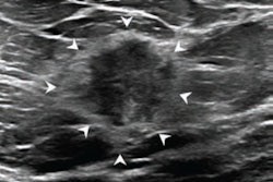
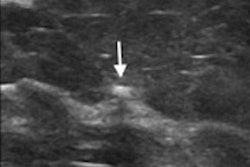
 Images from a 52-year-old woman who underwent prior breast ultrasound due to MRI abnormality show a suspicious breast lesion measuring 1.3 cm. Subsequent ultrasound was performed by a radiologist without breast ultrasound expertise for investigational purposes. A) Long-axis gray-scale ultrasound imaging shows a hypoechoic lesion. The lesion was classified by the study radiologist as BI-RADS category 4a. B) Long-axis greyscale ultrasound imaging shows the results of CAD analysis. CAD provided the result of "possibly benign" for both images. The lesion was therefore downgraded to category 3 based on CAD output. The pathologic assessment based on surgical resection was fibroadenoma. Images and caption courtesy of the American Journal of Roentgenology.
Cui and colleagues found that CAD downgraded 79.1% of BI-RADS 4a lesions to BI-RADS 3 and upgraded 6% of BI-RADS 3 lesions to BI-RADS 4a.
The research group also reported that the reading radiologists had significantly better diagnostic performance in several areas after the CAD software was introduced.
| Comparison of radiologist performance before, after CAD application | |||
| Measure | Without CAD | With CAD | p-value |
| Accuracy | 62.6% | 86.6% | < 0.001 |
| Sensitivity | 97.1% | 94.1% | 0.38 |
| Specificity | 46.0% | 82.9% | < 0.001 |
| Positive predictive value | 46.5% | 72.7% | < 0.001 |
| Negative predictive value | 97% | 96.7% | > 0.99 |
The team suggested that while CAD results must be integrated with clinical information and results of other breast imaging exams, the study findings point to the software's ability to improve patient care in settings where experienced breast radiologists may be in short supply.
"This [shortage] situation creates a need for additional tools to assist such radiologists in these efforts," the study authors wrote.