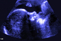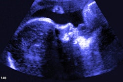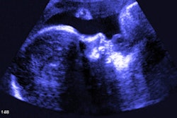
Routine third-trimester ultrasound leads to an over-70% reduction of undetected breech pregnancies along with improved neonatal outcomes, according to research published April 6 in PloS Medicine.
A team led by Dr. Asma Khalil from St. George's University Hospitals in London found that using standard ultrasound or point-of-care ultrasound (POCUS) can dramatically reduce all-term breech presentations that were previously undiagnosed.
"Our findings imply that a policy of either a third-trimester ultrasound by sonographers or POCUS by trained midwives was effective in reducing the proportion of all term breech presentations at the time of birth that were undiagnosed and associated neonatal complications," Khalil and colleagues wrote.
Accurate knowledge of fetal presentation is crucial for best-care practices during pregnancy and birth. Vaginal breech delivery, in which a fetus' buttocks, feet, or both are in position to come out first rather than the head, is tied to poor maternal and perinatal outcomes. While third-trimester ultrasound has been used for assessing fetal presentation, the researchers noted a lack of data on its impact on neonatal outcomes.
Khalil and co-authors wanted to test ultrasound's potential to reduce breech births. The team gathered data from St. George's and Norfolk and Norwich University Hospitals.
Before and after the implementation of routine third-trimester ultrasound scan or POCUS, there were 16,777 and 7,351 births in St. George's and 5,119 and 4,575 in Norfolk and Norwich, respectively. The team reported rates of breech presentation in labor across all groups of 3% to 4%.
Khalil and colleagues found that the percentage of all-term breech presentations that were undiagnosed significantly reduced after implementation of universal third-trimester ultrasound screening.
| Comparison of undiagnosed breech presentations before, after implementation of third-trimester ultrasound screening | ||||
| St. George's | Norfolk and Norwich | |||
| Before | After | Before | After | |
| Percentage of undiagnosed breech presentations | 14.2% | 2.8% | 16.2% | 3.5% |
The researchers also learned that the rate of undiagnosed breech was 71% lower after the implementation of a third-trimester ultrasound protocol. The team also found a moderate to high probability of reduction of hypoxic ischemic encephalopathy and extended perinatal mortality rates as the result of imaging women with ultrasound in the third trimester of pregnancy.
Using priors from a similar previous study, the investigators showed that the proportion of all-term breech presentations that were undiagnosed was 69% lower after POCUS was implemented.
The cost-effectiveness of POCUS should be explored to determine how feasible the technology's implementation could be on a wider scale for assessing fetal presentation, the study authors urged.


![Examples of ultrasound findings and techniques. (A) Images in a 39-year-old male patient with a mass in the left thigh. The mass is heterogeneous on the B-mode US image (compared with the patient in D) and showed increased microvascularity (superb microvascular imaging [SMI]) and shear-wave elastography (SWE) values. Undifferentiated pleomorphic sarcoma was diagnosed at biopsy (with pleomorphic rhabdomyosarcoma in surgical specimen). (B) Images in an 18-year-old male patient with a mass in the left leg. The mass is hypoechoic on the B-mode image, with no other findings suggestive of malignancy. The lesion is in contact with the cortex of the tibia, which is slightly irregular. CT revealed a doubtful anteromedial tibial erosion. The microvascular study demonstrated high vascularization, suggestive of malignancy. Periosteal Ewing sarcoma was diagnosed with both histologic and immunohistochemical confirmation. (C) Images in a 69-year-old female patient with a lump growing on the outside of the left leg. Multiple SWE examinations were performed (please note the high values obtained in the measurements, whereas the color map highlights the stiffness relative to adjacent tissues). SMI showed areas of increased vascularization to target for sampling. Undifferentiated spindle cell sarcoma was diagnosed at biopsy, with residual leiomyosarcoma in the surgical specimen after neoadjuvant therapy. (D) Images in a 56-year-old female patient with a mass in the right thigh. The mass is heterogeneous at both B-mode ultrasound (similar to patient A) and MRI (coronal T2-weighted spectral attenuated inversion recovery [SPAIR]; T1-weighted pre-contrast and postcontrast imaging), which even shows uptake after the administration of paramagnetic contrast material, which is traditionally suggestive of malignancy. Low values at SMI and elastography are suggestive of benignity. Spindle cell lipoma was diagnosed at biopsy, with atypical spindle cell lipomatous tumor in the surgical specimen.](https://img.auntminnie.com/mindful/smg/workspaces/default/uploads/2026/08/images-radiol250278fig2.APCFLSvX6p.jpg?auto=format%2Ccompress&fit=crop&h=100&q=70&w=100)


![Examples of ultrasound findings and techniques. (A) Images in a 39-year-old male patient with a mass in the left thigh. The mass is heterogeneous on the B-mode US image (compared with the patient in D) and showed increased microvascularity (superb microvascular imaging [SMI]) and shear-wave elastography (SWE) values. Undifferentiated pleomorphic sarcoma was diagnosed at biopsy (with pleomorphic rhabdomyosarcoma in surgical specimen). (B) Images in an 18-year-old male patient with a mass in the left leg. The mass is hypoechoic on the B-mode image, with no other findings suggestive of malignancy. The lesion is in contact with the cortex of the tibia, which is slightly irregular. CT revealed a doubtful anteromedial tibial erosion. The microvascular study demonstrated high vascularization, suggestive of malignancy. Periosteal Ewing sarcoma was diagnosed with both histologic and immunohistochemical confirmation. (C) Images in a 69-year-old female patient with a lump growing on the outside of the left leg. Multiple SWE examinations were performed (please note the high values obtained in the measurements, whereas the color map highlights the stiffness relative to adjacent tissues). SMI showed areas of increased vascularization to target for sampling. Undifferentiated spindle cell sarcoma was diagnosed at biopsy, with residual leiomyosarcoma in the surgical specimen after neoadjuvant therapy. (D) Images in a 56-year-old female patient with a mass in the right thigh. The mass is heterogeneous at both B-mode ultrasound (similar to patient A) and MRI (coronal T2-weighted spectral attenuated inversion recovery [SPAIR]; T1-weighted pre-contrast and postcontrast imaging), which even shows uptake after the administration of paramagnetic contrast material, which is traditionally suggestive of malignancy. Low values at SMI and elastography are suggestive of benignity. Spindle cell lipoma was diagnosed at biopsy, with atypical spindle cell lipomatous tumor in the surgical specimen.](https://img.auntminnie.com/mindful/smg/workspaces/default/uploads/2026/08/images-radiol250278fig2.APCFLSvX6p.jpg?auto=format%2Ccompress&fit=crop&h=112&q=70&w=112)