Ultrasonographic measurements of fetal nuchal translucency can lead to better management of chromosomal anomalies, according to a study published March 26 in JAMA Network Open.
Researchers led by doctoral candidate Kara Bellai-Dussault from the University of Ottawa in Ontario, Canada, found that pregnancies with nuchal translucency measurements greater than 2 mm are at increased risk of chromosomal anomalies. They highlighted that this means the widely used threshold of 3.5 mm may need to be reexamined.
“The findings were consistent through several sensitivity analyses,” Bellai-Dussault and colleagues wrote.
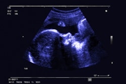
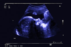
Fetal nuchal translucency is an ultrasound exam performed in the first trimester of pregnancy. It helps determine the risk of congenital conditions like Down syndrome being developed in fetuses. Specifically, it screens for trisomies 21 and 18 and other conditions.
A cutoff of 3.5 mm or greater is used as a marker for common and uncommon aneuploidies (i.e., missing chromosomes) as well as for a wide variety of genetic syndromes and structural anomalies. With this cutoff point, follow-up exams such as prenatal cell-free DNA screening or cytogenetic testing are recommended.
However, previous studies suggest ties between chromosomal anomalies and cutoff levels less than 3.5 mm. The Bellai-Dussault team evaluated the association between different nuchal translucency measurements and cytogenetic outcomes on a population level.
The study included data from 414,268 pregnancies in the Better Outcomes Registry & Network, the perinatal registry for Ontario, Canada. This included singleton pregnancies with an estimated date of delivery from September 2016 to March 2021.
The average maternal age at the estimated delivery date was 31.5 years. Also, of the total pregnancies, 359,807 (86.9%) had a nuchal translucency less than 2 mm. The researchers reported that the prevalence of chromosomal anomalies in this group was 0.5%.
The team found ties between increasing nuchal translucency measurements and an increased risk of chromosomal anomalies. This included an adjusted risk ratio (ARR) of 20.33 and an adjusted risk difference (ARD) of 9.94% for pregnancies with measurements of 3 mm to less than 3.5 mm.
Additionally, the ARR was 4.97 and the ARD was 1.4% when including only chromosomal anomalies beyond the commonly screened aneuploidies. This means excluding trisomies 21, 18, and 13 and sex chromosome aneuploidies.
The study authors highlighted that their results have important policy and quality assurance implications. This is because in some jurisdictions, prenatal cell-free DNA screening is offered after identifying increased nuchal translucency measures, while in others, such screening is part of first-tier prenatal screening.
“Screening programs should consider the value of nuchal translucency measurements when making decisions about whether to replace such measurements with [cell-free] DNA screening alone and may wish to reexamine the threshold of nuchal translucency at which diagnostic investigations are offered,” they wrote.
Bellai-Dussault's team also added that these findings highlight the importance of a robust quality assurance program to support nuchal translucency measurement, as “chronic undermeasurement can reduce the screening sensitivity.”
Finally, the authors called for future research to examine economic modeling to estimate benefits and costs to inform the best options for policy and practice.
The full study can be found here.