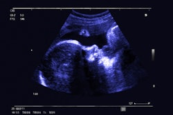
A novel method using ultrasound videos predicts fetal birth weight, according to Dutch research published October 20 in Computers in Biology and Medicine.
A team led by Szymon Płotka from the University of Amsterdam found that their method, which uses tabular clinical data to evaluate features in fetal ultrasound video scans, outperforms current algorithms and has comparable performance to that of clinicians.
“Our method has the potential to be applied in the clinical environment to assist in the selection of the safest type of delivery for both the mother and the child,” Płotka and co-authors wrote.
While ultrasound is the gold standard for assessing fetal growth and development, the modality is suspect to user dependence, which could cause variability in fetal biometric measurements. Accurate measuring of the head circumference, biparietal diameter, abdominal circumference, and femur length to predict fetal birth weight is important for proper pregnancy and delivery management.
The Płotka team previously described their automated fetal birth weight prediction method, which uses multimodal data and visual data processing, named BabyNet. BabyNet is a hybrid model that combines transformers and convolutional neural networks. It extends the 3D ResNet-18 architecture with a residual transformer module.
For the current study, the researchers wanted to test the performance of a refined version of BabyNet, which now includes a dynamic affine feature map transform module. This module uses tabular clinical data to improve fetal birth weight estimation. The researchers used fetal ultrasound video scans conducted within 24 hours before delivery along with relevant clinical indicators to estimate fetal birth weight. They also used the actual birth weight after delivery as the ground truth.
The team performed the development and evaluation of the latest version of BabyNet on a clinical set consisting of 582 2D fetal ultrasound videos and clinical records of pregnancies from 194 patients performed less than 24 hours predelivery.
It found that in fivefold cross-validation, BabyNet showed superior performance to that of clinicians and the original version of BabyNet. This included having lower error rates.
| |
BabyNet (original) |
Clinicians |
BabyNet (refined) |
|---|---|---|---|
| Mean absolute error (in grams) |
285 |
188 |
179 |
| Root mean squared error (in grams) |
374 |
238 |
203 |
| Mean absolute percentage error |
8.5% |
5.4% |
5.1% |
Furthermore, the refined version of BabyNet outperformed six other algorithms measuring fetal birth weight prediction.
In multicenter analysis, where BabyNet’s performance was compared to that of clinicians from four centers, the researchers found that BabyNet had significantly higher performance in all but one center (p = 0.09 for the one center).
Finally, the team reported that BabyNet showed the highest accuracy when measuring fetal abdominal ultrasound video scans. This included a mean absolute value of 175 grams, a root mean squared error of 200 grams, and a mean absolute percentage error of 5%.
The study authors wrote that using video analysis in fetal ultrasound exams has “several” advantages over static images. This includes using 2D spatiotemporal feature representations to improve performance.
“The utilization of short video sequences in our method might obviate the need for expert knowledge and skills for the precise estimation of fetal birth weight, as it did not fully depend on a reference standard plane,” they wrote.
The team called for future studies to have larger datasets with more operators from different skill groups and more scans per operator.
The full study can be found here.



![Examples of ultrasound findings and techniques. (A) Images in a 39-year-old male patient with a mass in the left thigh. The mass is heterogeneous on the B-mode US image (compared with the patient in D) and showed increased microvascularity (superb microvascular imaging [SMI]) and shear-wave elastography (SWE) values. Undifferentiated pleomorphic sarcoma was diagnosed at biopsy (with pleomorphic rhabdomyosarcoma in surgical specimen). (B) Images in an 18-year-old male patient with a mass in the left leg. The mass is hypoechoic on the B-mode image, with no other findings suggestive of malignancy. The lesion is in contact with the cortex of the tibia, which is slightly irregular. CT revealed a doubtful anteromedial tibial erosion. The microvascular study demonstrated high vascularization, suggestive of malignancy. Periosteal Ewing sarcoma was diagnosed with both histologic and immunohistochemical confirmation. (C) Images in a 69-year-old female patient with a lump growing on the outside of the left leg. Multiple SWE examinations were performed (please note the high values obtained in the measurements, whereas the color map highlights the stiffness relative to adjacent tissues). SMI showed areas of increased vascularization to target for sampling. Undifferentiated spindle cell sarcoma was diagnosed at biopsy, with residual leiomyosarcoma in the surgical specimen after neoadjuvant therapy. (D) Images in a 56-year-old female patient with a mass in the right thigh. The mass is heterogeneous at both B-mode ultrasound (similar to patient A) and MRI (coronal T2-weighted spectral attenuated inversion recovery [SPAIR]; T1-weighted pre-contrast and postcontrast imaging), which even shows uptake after the administration of paramagnetic contrast material, which is traditionally suggestive of malignancy. Low values at SMI and elastography are suggestive of benignity. Spindle cell lipoma was diagnosed at biopsy, with atypical spindle cell lipomatous tumor in the surgical specimen.](https://img.auntminnie.com/mindful/smg/workspaces/default/uploads/2026/08/images-radiol250278fig2.APCFLSvX6p.jpg?auto=format%2Ccompress&fit=crop&h=100&q=70&w=100)


![Examples of ultrasound findings and techniques. (A) Images in a 39-year-old male patient with a mass in the left thigh. The mass is heterogeneous on the B-mode US image (compared with the patient in D) and showed increased microvascularity (superb microvascular imaging [SMI]) and shear-wave elastography (SWE) values. Undifferentiated pleomorphic sarcoma was diagnosed at biopsy (with pleomorphic rhabdomyosarcoma in surgical specimen). (B) Images in an 18-year-old male patient with a mass in the left leg. The mass is hypoechoic on the B-mode image, with no other findings suggestive of malignancy. The lesion is in contact with the cortex of the tibia, which is slightly irregular. CT revealed a doubtful anteromedial tibial erosion. The microvascular study demonstrated high vascularization, suggestive of malignancy. Periosteal Ewing sarcoma was diagnosed with both histologic and immunohistochemical confirmation. (C) Images in a 69-year-old female patient with a lump growing on the outside of the left leg. Multiple SWE examinations were performed (please note the high values obtained in the measurements, whereas the color map highlights the stiffness relative to adjacent tissues). SMI showed areas of increased vascularization to target for sampling. Undifferentiated spindle cell sarcoma was diagnosed at biopsy, with residual leiomyosarcoma in the surgical specimen after neoadjuvant therapy. (D) Images in a 56-year-old female patient with a mass in the right thigh. The mass is heterogeneous at both B-mode ultrasound (similar to patient A) and MRI (coronal T2-weighted spectral attenuated inversion recovery [SPAIR]; T1-weighted pre-contrast and postcontrast imaging), which even shows uptake after the administration of paramagnetic contrast material, which is traditionally suggestive of malignancy. Low values at SMI and elastography are suggestive of benignity. Spindle cell lipoma was diagnosed at biopsy, with atypical spindle cell lipomatous tumor in the surgical specimen.](https://img.auntminnie.com/mindful/smg/workspaces/default/uploads/2026/08/images-radiol250278fig2.APCFLSvX6p.jpg?auto=format%2Ccompress&fit=crop&h=112&q=70&w=112)