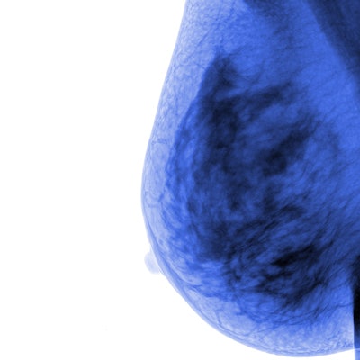
2D shear-wave elastography can spot differences between benign and malignant breast lesions, but lags behind strain elastography, according to a study published March 28 in the Journal of the American College of Radiology.
Researchers led by Aishwarya Pillai from Yale University found that elastography demonstrated high sensitivity and specificity when evaluating breast cancer and could prove beneficial in downgrading BI-RADS 4A or upgrading BI-RADS 3 lesions initially spotting on screening mammography.
"From a purely diagnostic standpoint, integration into current protocols for working up suspicious breast lesions may be considered," Pillai and colleagues wrote. "However ... we cannot recommend using shear-wave elastography to the exclusion of strain elastography at this time."
When lesions are found on mammography, they're assigned a BI-RADS score ranging from 0 to 6. BI-RADS 3 lesions are likely benign, with a less than 2% risk for malignancy. BI-RADS 4 lesions are trickier because they are categorized as suspicious. These lesions are divided into subcategories: 4A (2% to 10% chance of malignancy), 4B (10% to 50% chance of malignancy), and 4C (50% to 95% chance of malignancy). These often require biopsy for further workup.
The researchers wrote that more objective evaluation methods are needed though since BI-RADS 3 and 4 assessments can have low specificity. Previous research suggests that shear-wave elastography can characterize breast lesions with high sensitivity and specificity. This is because malignant lesions are often significantly stiffer than benign lesions.
Pillai et al wanted to look at 2D shear-wave elastography's performance in differentiating lesions as well as the feasibility of determining the optimum threshold parameter.
They included 87 prospective and retrospective studies published between 2010 and 2021 in their analysis. The studies involved a total of 17,810 women with an average age of 42.3 years with 19,043 lesions (7,623 malignant). They also used the average, maximum, minimum, or standard deviation of shear-wave speed for classification.
| Performance of shear-wave parameters for differentiating benign and malignant breast lesions | |||
| Sensitivity | Specificity | Areas under the receiver operating characteristic curve | |
| Summary | 0.86 | 0.87 | 0.93 |
| Maximum | 0.83 | 0.88 | 0.92 |
| Minimum | 0.86 | 0.81 | 0.90 |
| Standard deviation | 0.82 | 0.88 | 0.92 |
However, these results are not superior to strain elastography, which had a sensitivity of 96% and a specificity of 88% in a review published in 2018. The review also showed that strain elastography could downgrade BI-RADS 4B lesions to BI-RADS 3. Researchers wrote that shear-wave elastography can support only a downgrade from BI-RADS 4A to BI-RADS 3.
Still, the team said shear-wave elastography works well in evaluating benign lesions over strain elastography and the two methods could be combined to evaluate certain breast tumors.
They added that shear-wave elastography's role is not to replace biopsy altogether but rather serve as an adjunct to BI-RADS categorization and that their results can further discussions on definitive recommendations for society guidelines.
"Given the possibility that each method may be better for characterizing certain types of lesions, another important avenue for further study is determining whether combining elastographic methods will increase their diagnostic utility," the study authors wrote.
They also called for large, prospective multicenter trials to test the cutoff parameters, as well as alternative, nonstandard models for data pooling.




![Examples of ultrasound findings and techniques. (A) Images in a 39-year-old male patient with a mass in the left thigh. The mass is heterogeneous on the B-mode US image (compared with the patient in D) and showed increased microvascularity (superb microvascular imaging [SMI]) and shear-wave elastography (SWE) values. Undifferentiated pleomorphic sarcoma was diagnosed at biopsy (with pleomorphic rhabdomyosarcoma in surgical specimen). (B) Images in an 18-year-old male patient with a mass in the left leg. The mass is hypoechoic on the B-mode image, with no other findings suggestive of malignancy. The lesion is in contact with the cortex of the tibia, which is slightly irregular. CT revealed a doubtful anteromedial tibial erosion. The microvascular study demonstrated high vascularization, suggestive of malignancy. Periosteal Ewing sarcoma was diagnosed with both histologic and immunohistochemical confirmation. (C) Images in a 69-year-old female patient with a lump growing on the outside of the left leg. Multiple SWE examinations were performed (please note the high values obtained in the measurements, whereas the color map highlights the stiffness relative to adjacent tissues). SMI showed areas of increased vascularization to target for sampling. Undifferentiated spindle cell sarcoma was diagnosed at biopsy, with residual leiomyosarcoma in the surgical specimen after neoadjuvant therapy. (D) Images in a 56-year-old female patient with a mass in the right thigh. The mass is heterogeneous at both B-mode ultrasound (similar to patient A) and MRI (coronal T2-weighted spectral attenuated inversion recovery [SPAIR]; T1-weighted pre-contrast and postcontrast imaging), which even shows uptake after the administration of paramagnetic contrast material, which is traditionally suggestive of malignancy. Low values at SMI and elastography are suggestive of benignity. Spindle cell lipoma was diagnosed at biopsy, with atypical spindle cell lipomatous tumor in the surgical specimen.](https://img.auntminnie.com/mindful/smg/workspaces/default/uploads/2026/08/images-radiol250278fig2.APCFLSvX6p.jpg?auto=format%2Ccompress&fit=crop&h=100&q=70&w=100)


![Examples of ultrasound findings and techniques. (A) Images in a 39-year-old male patient with a mass in the left thigh. The mass is heterogeneous on the B-mode US image (compared with the patient in D) and showed increased microvascularity (superb microvascular imaging [SMI]) and shear-wave elastography (SWE) values. Undifferentiated pleomorphic sarcoma was diagnosed at biopsy (with pleomorphic rhabdomyosarcoma in surgical specimen). (B) Images in an 18-year-old male patient with a mass in the left leg. The mass is hypoechoic on the B-mode image, with no other findings suggestive of malignancy. The lesion is in contact with the cortex of the tibia, which is slightly irregular. CT revealed a doubtful anteromedial tibial erosion. The microvascular study demonstrated high vascularization, suggestive of malignancy. Periosteal Ewing sarcoma was diagnosed with both histologic and immunohistochemical confirmation. (C) Images in a 69-year-old female patient with a lump growing on the outside of the left leg. Multiple SWE examinations were performed (please note the high values obtained in the measurements, whereas the color map highlights the stiffness relative to adjacent tissues). SMI showed areas of increased vascularization to target for sampling. Undifferentiated spindle cell sarcoma was diagnosed at biopsy, with residual leiomyosarcoma in the surgical specimen after neoadjuvant therapy. (D) Images in a 56-year-old female patient with a mass in the right thigh. The mass is heterogeneous at both B-mode ultrasound (similar to patient A) and MRI (coronal T2-weighted spectral attenuated inversion recovery [SPAIR]; T1-weighted pre-contrast and postcontrast imaging), which even shows uptake after the administration of paramagnetic contrast material, which is traditionally suggestive of malignancy. Low values at SMI and elastography are suggestive of benignity. Spindle cell lipoma was diagnosed at biopsy, with atypical spindle cell lipomatous tumor in the surgical specimen.](https://img.auntminnie.com/mindful/smg/workspaces/default/uploads/2026/08/images-radiol250278fig2.APCFLSvX6p.jpg?auto=format%2Ccompress&fit=crop&h=112&q=70&w=112)